Biosorption of Water Pollutants by Fungal Pellets
by
 , and
, and
Adriana Jazmín Legorreta-Castañeda
, 
Carlos Alexander Lucho-Constantino
,
Rosa Icela Beltrán-Hernández
,
Claudia Coronel-Olivares
and
Gabriela A. Vázquez-Rodríguez
*
Área Académica de Química, Universidad Autónoma del Estado de Hidalgo, Carr. Pachuca-Tulancingo Km. 4.5, Pachuca C. P. 42184, Mexico
*
Author to whom correspondence should be addressed.
Water 2020, 12(4), 1155; https://doi.org/10.3390/w12041155
Submission received: 15 March 2020
/
Revised: 12 April 2020
/
Accepted: 14 April 2020
/
Published: 17 April 2020
(This article belongs to the Special Issue Microbial Action in Wastewater and Sludge)
Abstract
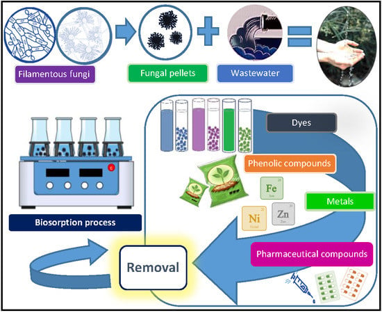
:Fungal biosorption is an environmental biotechnology based on the ability of the fungal cell wall to concentrate harmful water pollutants. Among its advantages are its simplicity, high efficiency, flexibility of operation, and low cost. The biosorptive performance of fungal pellets is getting growing attention since they offer process advantages over the culture of disperse mycelia, such as an enhanced biomass separation, and a high resilience in severe environmental conditions. In this review, biosorption capacity of fungal pellets towards heavy metals, dyes, phenolic compounds, humic substances, pesticides, and pharmaceuticals was reviewed. Available data about the adsorption capacity of pellets, their removal efficiency, and the operational conditions used were collected and synthesized. The studies relying on biodegradation were discarded to present only the possibilities of fungal pellets for removing these concern pollutants through biosorption. It was found that the biosorption of complex mixtures of pollutants on fungal pellets is scarcely studied, as well as the interfering effect of anions commonly found in water and wastewater. Furthermore, there is a lack of research with real wastewater and at pilot and large scale. These topics need to be further explored to take full advantage of fungal pellets on improving the quality of aquatic systems.
1. Introduction
A growing number of pollutants are discharged in the aquatic environment through industrial and municipal wastewater streams. In these, pollutants so diverse as heavy metals, dyes, pesticides, and pharmaceutical compounds, among others, are commonly found in a wide range of concentrations [1,2,3,4]. Some of these pollutants are highly persistent and can adversely impact the aquatic communities for a long time. For instance, dyes inhibit the aquatic life necessary to the natural attenuation of aquatic pollution, because they diminish primary productivity by reducing the entrance of solar light [2], while some pharmaceutical products might spread antibiotic resistance or endocrine disruption phenomena [5]. For these reasons, the dispersal of the aforementioned pollutants must be prevented through adequate wastewater treatment systems.
Adsorption, usually on activated carbon, has demonstrated to be a well-suited technology for the removal of persistent pollutants otherwise difficult to eliminate from wastewater. This is due to the versatility, efficiency, and low cost of activated carbon [6]. Yet there are some drawbacks associated with the use of this sorbent, the main of which is the need of chemical or thermal regeneration. This process can be impractical and expensive at full scale, produces additional pollution, and causes a considerable loss of the original material.
Biosorption, the removal of pollutants by adsorption on biological material, has emerged as an attractive technology due to its simplicity, high efficiency, flexibility of operation, and low cost [7,8,9]. Other assets of biosorption are the possibility of low-cost regeneration of the biosorbent and the recovery of the sorbate [6,10,11].
A wide range of natural materials and agricultural or industrial waste has been used as biosorbents [12,13]. This includes microbial biomass in various life stages (in lag, log, or stationary phases) or even dead. In some cases, dead microbial biomass is treated to enhance its surface properties, which are due to cell components such as alginic acid, chitin or cellulose [10,14]. Seaweeds, fermentation waste and out-purged activated sludge are among the most used biosorbents [6,13].
Although the biosorption term is widely used to denote the adsorption processes carried out onto any biological material, in this review a narrower scope was preferred. Biosorption was considered as the active or passive capture of organic and inorganic species from aqueous solutions by microbial biomass, which allows the removal (and even the further recovery) of these pollutants in an environmentally-sound way [10]. This term includes mechanisms depending on metabolism, but also independent of it, such as physical and chemical adsorption, ionic exchange, complexation, chelation, and microprecipitation, which occur mainly on the cell surface [13].
Lately, fungal biosorption has received substantial attention as well, because it is considered as an eco-friendly, economical, and effective alternative for the removal of pollutants from wastewater [15]. Fungi constitute a group of both unicellular and multicellular organisms with outstanding industrial and environmental applications. Multicellular fungi display development mechanisms quite different from those shown by plants and animals; these mechanisms are characterized by the formation of filament assemblies called hyphae, which grow only apically [16]. Fungal filaments contain all the components of eukaryotic cells and are covered by a unique cell wall with prominent amounts of glycoproteins, chitin, and glycans. Functional groups allowing biosorption such as hydroxyl, amine, carboxyl, among others, are profusely present in fungal cell walls [8]. Given the fact that waste fungal biomass is generated abundantly from several industrial processes, it constitutes also a promising source of biosorbents [14,17].
In liquid media, filamentous fungi grow as disperse mycelium or form granules visible as microspheres, known as fungal pellets. Considerable research has been conducted recently to explore the potential of fungal pellets in industrial processes, as well as the factors influencing the granulation mechanism [2,7]. These pellets present several advantages over disperse mycelia, such as lower viscosity of the culture medium and easier biomass separation [7]. Besides, fungal pellets have proved to be resilient towards severe conditions, such as acidic media, fluctuating inputs of toxicants or low concentrations of nutrients [18], which is advantageous in environmental processes dealing with industrial wastewater or acid mine drainage, for instance. Furthermore, fungal pellets are a well-suited source of enzymes useful for organic compounds degradation [7]. Several factors have been reported to be crucial to culturing fungal pellets. Among them, pH, agitation, medium composition, inoculation mode, and additives are commonly mentioned [19]. However, fungal pellets can also show some drawbacks, as hindered internal transport of nutrients leads to the apparition of non-viable zones inside the granules [7].
The aim of this paper is to review the most common applications of fungal pellets to the removal of water pollutants through biosorption. We revised the scientific literature presenting the use of fungal pellets for eliminating heavy metals, dyes, phenolic compounds, pesticides, humic substances, and pharmaceutical compounds, and we collected the data related to the adsorption capacity of pellets, their removal efficiency, and the operational conditions used. Some of the reviewed compounds are prone to be degraded through oxidative metabolic pathways [20]; however, we discarded the experimental data relying on biodegradation, to present only the possibilities of fungal pellets for removing these concern pollutants through biosorption. Similarly, we discarded the biosorption studies dealing with fungal pellets but that had been treated in such a way that the granular shape was shattered (i.e., by grinding) before the biosorption tests.
2. Biosorption of Some Relevant Water Pollutants by Fungal Pellets
Either alive or dead, fungal biomass has demonstrated to be an efficient biosorbent, mostly due to several functional groups found in the cell wall [8,15]. However, the biosorption studies have focused mostly on dead fungal cells, because they offer some advantages over live cells. First, the medium requirements, as of pH value, contents of nutrients, or temperature, are more stringent for the biosorption carried out by live cells. By using dead biomass, the addition of growth nutrients is avoided. Second, dead fungal biomass can be stored easily for longer periods [21]. Biosorption on dead biomass depends on the chemical nature of the adsorbate (as its molecular size or ionic charge), the type of biomass and its chemical treatment, as well as the environmental conditions of the process [22,23].
In the last years, the biosorption of dyes, phenolic compounds, pesticides, and heavy metals, among other pollutants of concern, has been studied in several fungal species, by using dead or alive cells and a wide array of physicochemical pretreatments. These studies are described in more detail in the following sections, after a short presentation of the pollutants considered.
2.1. Heavy Metals
The mobilization of heavy metals through the extraction of minerals and their further processing has led to the broad dispersion of these pollutants [24]. Thus, large wastewater volumes are generated annually with varying levels of heavy metals; among them, Cd, Cr, Cu, As, Hg, and Pb can be highlighted due to their toxicity and ubiquity [25,26].
There are two main sources of heavy metals in wastewater effluents: natural and anthropogenic. The first includes soil erosion, volcanic activities, weathering of rocks and minerals [25], while the main anthropogenic sources are paints and pigments, plastic stabilizers, electroplating, incineration of cadmium-containing plastics, and phosphate fertilizers for Cd [27,28]; tanneries and steel industries for Cr [29]; pesticides and wood preservatives for Cu [29,30] and As [31]; release from Au-Ag mining and coal combustion, and medical waste for Hg [32,33]; industrial effluents, kitchen appliances, surgical instruments, steel alloys, automobile batteries [34], aerial emission from combustion of leaded petrol, battery manufacture, herbicides, and insecticides for Pb [27,31,32].
The presence of heavy metals in wastewater raises several environmental issues, because these are conservative pollutants to which the “biodegradability” term does not apply. In addition, they are mobile and toxic in aquatic ecosystems [24,25,26,27]. Other of the main concerns is their potential to accumulate in living organisms, and then to biomagnificate through the trophic chains (i.e., the organisms from the higher trophic levels are polluted with higher contents of heavy metals). In this way, human health is threatened if these pollutants are present at high concentrations in food and water [35,36]. Besides, heavy metals cause oxidative stress by means of the formation of free radicals [37]. Oxidative stress refers to the enhanced generation of reactive oxygen species that may overwhelm the antioxidant defenses of cells, resulting in their permanent damage or death [38,39].
In view of the above, it is imperative to remove the heavy metals from wastewater before their discharge to the aquatic environment. Chemical precipitation, oxidation or reduction, ionic exchange, electrochemical processes, reverse osmosis, and other membrane separation technologies are among the most used treatment methods [26,40,41,42]. These technologies present some drawbacks, such as their relatively high cost, in some cases derived from the constant inputs of chemical reagents or energy, or their inefficiency to treat diluted streams (with concentrations of metals below 100 mg·L−1) [43]. This has generated a considerable interest in biosorption, which has demonstrated its efficiency to remove heavy metals at low operating costs [44,45,46]. In particular, filamentous fungi biomass has the potential to do so [47,48]. Several types of fungal biomass are promising sources of biosorbents to remove heavy metals from aquatic streams. These sources must be available at reduced costs, be able to remove high amounts of metals and, if possible, to be regenerated and reused more than once. Such a source could arise from the waste of large-scale fungal bioprocesses, which includes the antibiotic industrial production.
It is worth mentioning that, unlike other pollutants, heavy metals can be removed from wastewater by a biosorbent through different mechanisms such as: (i) chemical transformations involving phase changes (i.e., redox reactions or alkylation), (ii) bioaccumulation, which includes metabolism-dependent processes leading to the metal transport into the fungal cells, and (iii) biosorption, which is a surface mechanism that does not involve any metabolic process. The latter mechanism is considered to be the most significant in metals removal by fungal biomass, which can be attributed to ion exchange, coordination or covalent bonding to the cell wall [7]. On live fungal biomass, the sorption of heavy metals involves two stages: a fast metabolism-independent phase that relies on the available surface and it is of physicochemical nature, followed by a slow metabolism-dependent phase that implies the ion transport across the cell membrane [49,50]. From a quantitative point of view, the surface adsorption can represent the most part of the total ion removal, so that the union to the cell membranes could be the most significant mechanism of metal removal. This mechanism occurs in both alive and dead fungal biomass. In fact, dead cells can adsorb some metallic ions in a greater extent than live cells [50,51].
Rather than focusing separately on the fungal biosorption of each heavy metal reported in the bibliography, this section will review the main parameters involved in metal uptake by fungal biomass, which are the solution pH, the initial metal concentration, the biomass pretreatment, and the evaluation of the metal removal in multicomponent systems. Table 1 presents the adsorption capacities measured in several published studies, as well as the used experimental conditions.
2.1.1. Effect of pH on Biosorption of Metals
As we have mentioned in previous sections, the solution pH affects the speciation of metal ions, and also influences the surface properties of the sorbents [52,53], affecting the dissociation of binding sites and their surface charge, the activity of functional groups (mainly carboxylate, phosphate, and amino groups) in the biomass [54], as well as the competition of metallic ions with hydronium ions for the adsorption sites [53].
The effect of pH in biosorption processes depends on the strain and the metal ion to be removed. Binupriya et al. [55] evaluated the biosorption of metal ions in aqueous solutions with pellets of Aspergillus japonicus; in the case of Ni(II), the removal increased as the pH increased from 2 to 10, reaching a maximum removal of 100% at pH 9. In the case of Fe(II), the removal improved as the pH increased from 2 to 6; at pH 6, the removal was 100%, and when the pH was extended above this value and until pH 10, the removal remained higher than 90%. This trend has also been reported for Zn removal by Phanerochaete chrysosporium pellets [56], as the adsorption capacities increased as the pH values rose from 2 to 7. The maximum removal efficiencies (48.6 ± 4.5%) were observed at pH 7.0, which was attributed to the dissociation of protons from the carboxylic acid group, one of the major components of the fungal cell wall. As the pH of the aqueous solution increased, the dissociation of protons also augmented, as well as the presence of negatively-charged groups in fungal surface and therefore the number of binding sites available for complexation of metal cations [56]. Concerning the removal of U(VI) with Funalia trogii pellets, the optimum pH was 5, which was attributed to the presence of ligands such as amino, carboxyl, and phosphate groups on the surface of the pellets at this pH value [57]. The authors indicated that the decrease in the amount of metal absorbed at pH higher than 5 was due to the formation of uranyl carbonate complexes [57]. Concerning the biosorption of Cu, it has been reported that when pH value is higher than 5, the adsorption on fungal pellets decreases due to the precipitation of copper hydroxide [58,59]. As most metals complex and precipitate at alkaline pH values, biosorption is usually evaluated in a 2–7 pH range to attribute the metal removal only to the biosorption process.
On the other hand, at very acid pH values, low metal adsorption capacities have been reported, which is mainly attributed to the hydronium ions competing with the metal ions for the binding sites. For instance, the removal of Cu, Ni, and Zn is negligible at pH 3, due to the effects of the competition between the cations and the hydronium ions for the biosorption sites in Aspergillus niger pellets [53]. In the case of Cu, a low biosorption capacity was observed at pH values below 3.0 when alkali-pretreated and viable pellets of Rhizopus oryzae were tested [1]. In column experiments, Gabriel et al. [58] reported a low Cu biosorption capacity with pellets of Pleurotus ostreatus at pH 3 (1.92 mg·g−1) compared to the value obtained at pH 4 (7.92 mg·g−1). Many authors agree that the pH optimum for Cu biosorption is at 4–5 pH range [1,53,61]. Mishra and Malik [61] obtained the best metal uptake yields (79.8% and 77.2%) at pH 4 and 5, respectively, with Aspergillus lentulus pellets. For Rhizopus oryzae pellets, the maximum Cu adsorption capacities (52.91–61.73 mg·g−1) were reported at pH 4 [59]. In another study, the maximum Cu removal with alive pellets of Phanerochaete chrysosporium (50%) and with Funalia trogii pellets (61%) was obtained at pH 5 [63].
In the case of Cr(VI) biosorption, the removal by Aspergillus japonicus pellets decreased from 100 to 40% as the pH value increased from 2 to 10 [55]. Filipović-Kovačević et al. [53] reported that the biosorption of Cr(VI) on Aspergillus niger pellets followed this same trend, since the maximum capacity of adsorption was obtained at pH 2 and then it decreased as the pH increased from 2 to 7. This was explained by the speciation of Cr(VI), which predominates as HCrO4− at concentrations less than 500 mg·L−1 and low pH values, although another species as Cr2O72−, Cr3O102−, and Cr4O132− coexist in acid media [75]. As all these Cr(VI) species are negatively charged, a decrease in pH leads to a higher protonation of the fungal surface, creating a stronger attraction between the adsorbate and the biosorbent. Therefore, as the pH increases, the surface charge of the fungal pellets becomes negative, leading to weak bondings with the negatively-charged Cr species. Besides, in alkaline environments other negative ions such as OH- are likely to compete with the anion predominant at higher pH values (CrO42−) for the biosorption on the fungal biomass [75,76].
Finally, several works report a negligible effect of pH in the biosorption process. It is the case of As(III), for which almost complete removal was obtained at several concentrations of both Phanerochaete chrysosporium pellets and adsorbate, while a negligible effect of pH values comprised between 5 and 9 was observed [66]. In preliminary experiments with Pb(II) and Aspergillus lentulus pellets, Mishra and Malik [61] concluded that pH did not significantly alter the metal uptake.
2.1.2. Effect of Initial Metal Concentration on Biosorption
Another important parameter in the biosorption of metals with fungal pellets is the initial metal concentration in the solution. On the one hand, if the metal biosorption and bioaccumulation of the metal are being evaluated in live pellets, its initial concentration can affect the growth of the pellets [61]. On the other hand, the initial concentration of metal ions in the solution plays a key role as a driving force to overcome the mass transfer resistance between the aqueous phase and the biosorbent [77].
Mishra and Malik [61] evaluated the growth of Aspergillus lentulus pellets in presence of Cu(II), Cr(III), Ni(II), and Pb(II). A lower biomass production was observed, compared to a control, when metals were added. The biomass growth reduced 19% in presence of 70 mg Ni2+·L−1; this reduction was of 76% when the metal concentration increased at 140 mg·L−1. In the same study, after five days of growth, the addition of 80 mg Cu2+·L−1 reduced 16% the pellets’ biomass, while this reduction was of 77% at 800 mg Cu2+·L−1.
The metal uptake capacity of Aspergillus lentulus was also evaluated as a function of the initial concentration of metal ions (Cu(II), Cr(III), Ni(II), and Pb(II)) in the medium [61]. The removal of Cu(II) and Ni(II) ions was enhanced by increasing the initial metal ion concentration. The maximum specific metal uptake was determined as 124.5 mg·g−1 (at 800 mg·L−1) for Cu(II), as 11 mg·g−1 (at 140 mg·L−1) for Ni(II), 331.5 mg·g−1 for Cr(III), and 1120.6 mg·g−1 for Pb(II) (at 4000 mg·L−1 for both metal ions) [61]. A high initial concentration provides an increased driving force to overcome all mass transfer resistance of metal ions between the aqueous and solid phase, resulting in a higher probability of collision between metal ions and biosorbents [61,77].
The biosorption capacity of Pb(II) (1120.6 mg·g−1) obtained with live Aspergillus lentulus pellets [61] is 70-fold higher than the values obtained with live pellets of Phanerochaete chrysosporium, namely 16 mg·g−1 [68], and around 120-fold higher than the capacity (9.0 mg·g−1) measured by Yetis et al. [69]. By using alkali-pretreated pellets of the same strain, low Pb(II) biosorption capacities were reported (15.2–23.7 mg·g−1 and 20.1–48.2 mg·g−1, respectively) [68,69]. This differences between adsorption capacities are mainly due to the initial metal concentration used (50 mg·L−1) in these works [68,69] compared to the (4000 mg·L−1) evaluated by Mishra and Malik [61].
An increase in the Ni concentration (from 100 to 500 mg·L−1) resulted in an approximately fivefold increase in the biosorption capacity of this metal onto Rhizopus arrhizus pellets from 61.2 to 348.8 mg·g−1. However, the biosorption capacity decreased when the nickel concentration exceeded 500 mg·L−1 [72].
Some general comments on the literature reviewed so far can be made. First of all, research work concerning the evaluation of fungal pellets for the biosorption of low concentrations of metal ions is scarce. The concentrations being evaluated should be as realistic as possible, and for some metals (i.e., precious metals or radionuclides) their concentrations in wastewater are hardly higher than 1–10 mg·L−1 [66]. Besides, other compounds that can intervene negatively in the biosorption processes of metals should be more explored, such as the most common anions present in groundwater and industrial effluents (i.e., chlorides, nitrates, fluorides, phosphates, and sulfates) [53]. In the Section 2.1.6, the few articles that have reported the interference of these anions and their competition with different metal ions will be discussed.
2.1.3. Effect of Diameter of Pellets on Biosorption of Metals
The pellet diameter determines the surface area of the biosorbent, which is a key factor in biosorption processes. It also determines the number of metal binding functional groups readily exposed to the metal ions in solution. Fu et al. [59] evaluated the effect of the diameter of the Rhizopus oryzae pellets on the removal of Cu(II) at pH 4 and an initial concentration of 100 mg·L−1. The adsorption capacity decreased from 37.1 to 18.4 mg·g−1 when the diameter of the pellet increased from 0.4 mm to 2.0 mm. This was attributed to a decrease in the surface area, which was responsible for a lower availability of exposed binding sites in biomass of larger diameter. When the diameter was higher than 1.2 mm, the autolysis of the pellets was likely to further decrease the availability of metal-binding functional groups.
Li et al. [67] evaluated the effect of pellets diameter (from 1.32–1.57 mm to 3.5–5.50 mm) on the Cd adsorption on Phanerochaete chrysosporium, at pH 4.5, 25 °C, and an initial metal concentration of 50 mg·L−1. The maximum Cd uptake (15.2 mg·g−1) was obtained when the pellet diameter was comprised between 1.58 and 2.03 mm. As for Fu et al. [59], the adsorption capacity decreased to 9.86 mg·g−1 when the diameter of the pellet increased to 3.5–5.5 mm.
Gabriel et al. [58] also studied the Cu biosorption by pellets of different strains, namely Aspergillus carbonarius, Lepista nuda, Oudemansiella mucida, Phanerochaete chrysosporium, Pleurotus ostreatus, and Pycnoporus cinnabarinus, among others. Although the variety of conditions at which the adsorption experiments were carried out hinders the comparisons, it is noteworthy that some of the reviewed papers about Cu biosorption report the same environmental conditions, i.e., pH 4, 25 °C, and a stirring rate of 50 rpm. In these conditions, the highest adsorption capacities were observed for the following fungal species: O. mucida (8.8 mg·g−1), L. nuda (6.3 mg·g−1), P. cinnabarinus (5.1 mg·g−1), and P. ostreatus (4.8 mg·g−1). The differences in these values were attributed to dissimilarities in the cell wall composition and to the physical properties of the pellets. From a biotechnological point of view, the fungal pellets of O. mucida, P. chrysosporium, and P. ostreatus were considered as having the best mechanical properties among the species studied [58]. These pellets had a size big enough for handling (3–7 mm) but at the same time with an adequate surface area. Thus, due to their high adsorption capacity, ease of handling, and resistance to mechanical disintegration, these fungal species were deemed well-suited to environmental applications.
2.1.4. Effect of Pretreatment of Fungal Pellets on Biosorption of Metals
It has been reported that the biosorption capacities of fungal pellets can improve with the modification of their surface area, either with acid [72], alkaline [69], or thermal pretreatments [52]. Other strategies are based on the chemical modification of the pellet surface with ligands having oxygen donor atoms, such as amidoxime [57], or the immobilization of nanoparticles of some metals on the fungal pellet surface [56,64]. Table 2 summarizes the results of some studies that have evaluated the pretreatment effect on the biosorption of metals.
Some of the reasons for using dead or inactive pellets in metal biosorption are that maintaining the viability of the microbial cells during the process requires a continuous supply of nutrients, and that avoiding toxicity against the cells might need a certain control of the metal concentration. Therefore, the use of non-living microbial cells can eliminate these problems, while enabling the biomass regeneration and reuse for several cycles [78].
Bayramoglu and Arica [52] evaluated the removal of Hg(II), Cd(II), and Zn(II) with alive (active) and dead (heat-inactivated) pellets of Lentinus edodes. The surface areas of the alive and heat-inactivated fungal pellets were measured by the BET method and were found to be 0.89 and 1.18 m2·g−1 of fungal biomass, respectively. The surface area in the native pellets was increased after thermal treatment, which enhanced the biosorption of the metals evaluated. So, the heat-inactivated pellets showed better adsorption capacities for Hg (419.1 mg·g−1), Cd (299.4 mg·g−1), and Zn (63.3 mg·g−1) compared to the alive fungal pellets (358.1, 86.4, and 37.7 mg·g−1, respectively).
The chemical modification with amidoxime appears to increase the biomass surface area and to favor the adsorption of heavy metals, as reported for Trametes trogii pellets evaluated for the biosorption of U(VI) [57]. Maximum biosorption capacities of modified and native pellets were found to be 447 and 238 mg g−1, respectively. This was explained by the fact that the amidoxime pretreatment increased 3.3-fold the surface area of fungal pellets, as well as the total available surface amino groups (2.54 mmol·g−1 of dry modified biomass against 0.36 mmol·g−1 dry native biomass).
One of the most recent proposals for improving metal removal is the synergy between biosorbents and nanomaterials [56]. Particles in the nano-size range possess altered properties compared to their bulk materials, including larger surface areas, higher reactivities, and faster adsorption kinetics [64], which makes them particularly attractive as sorbents [79]. Recently, fungal pellets have been proposed as biomass carriers of nanoparticles to make up new biocomposites for the treatment of wastewater. Abundant functional groups on the mycelium surface provide the feasible environment for the assembly and enhance the dispersibility of nanoparticles [64]. Phanerochaete chrysosporium pellets provided with nanosized Se showed to be better biosorbents as they removed more Zn (88.1 ± 5.3%) compared to Se- free fungal pellets (56.2 ± 2.8%) at pH 4.5 and an initial Zn concentration of 10 mg·L−1. This improvement in biosorption performance was attributed to a more negative surface charge density, and hence to a higher concentration of sorption sites [56].
Ding et al. [64] studied the immobilization of iron oxide nanoparticles in pellets of Penicillium sp., which showed the following advantages: the nano-Fe3O4 particles can uniformly grow on the surface of Penicillium sp. with no aggregation; Penicillium sp. can be used as the template to direct and control the structure of the nano-Fe3O4 from the micro-scale level; and fungal pellets are more environmentally-friendly and cost-effective than other reported templates. The sorption results for three radionuclides (Sr(II), Th(II), and U(VI)) on both the alive native pellets and the Fe3O4-pellets of Penicillium sp. are shown in Table 2. Yet the increase of sorption capacity for the composite fungus-Fe3O4 toward the radionuclides was not obvious, probably because some surface functional groups of Penicillium sp. were occupied by nano-Fe3O4, leading to a partial loss of the ability of binding with radionuclides.
2.1.5. Biosorption of Heavy Metals Form Mixtures
Little attention has so far been given to the multi-component biosorption of metal ions. However, these studies are more environmentally-relevant than those carried out with single metal ions, because they reflect more closely the state of the actual aquatic media. In these multi-systems, the biosorption depends, as shown above, on the biosorbent surface features, physicochemical parameters such as the solution pH, and the initial concentrations of both the adsorbate and the adsorbent, but also on the number and characteristics of the involved ions, among other factors [67]. Some relevant studies dealing with multi-metal ion biosorption will be discussed below.
Lead represents a serious and well-known environmental issue, because it induces dysfunctions in the neurologic, renal, and reproductive systems, particularly in young children. It is often released with cadmium from certain chemical processes and battery manufacturing [67,80]. Li et al. [67] studied the competitive biosorption of Cd2+ and Pb2+ by pellets of P. chrysosporium in the optimal physicochemical conditions determined for each metal separately. The comparison between the competitive biosorption of Cd2+ and Pb2+ showed that the biosorption of Pb2+ by P. chrysosporium pellets was preferential to that of Cd2+. Since both electronegativity and ionic radius of Pb2+ were larger than Cd2+, there might be a stronger chemical and physical affinity for Pb2+ on P. chrysosporium.
Bayranoglu et al. [52] evaluated the biosorption of cadmium, mercury, and zinc, because these three metals are commonly discharged together in wastewater by many industrial activities; however, cadmium and mercury raise the largest human health concerns. Cadmium is classed as a human and animal carcinogen (group 1) by the IARC (International Agency for Research on Cancer) [81]. As to mercury, it has been related to the induction of more than 250 symptoms [82]. The biosorption of the Cd2+, Hg2+, and Zn2+ multi-system was studied with dead and alive fungal pellets of Lentinus edodes [52]. For both types of pellets, and for all the metal ions (at individual concentrations varying from 25 to 600 mg·L−1), the highest adsorption capacity occurred at pH 6. Dead and alive pellets showed the same affinity order for the metal ions both individually and in mixture: Hg2+ > Cd2+ > Zn2+. In fact, the adsorption capacity of Hg2+ was one order of magnitude higher than the Zn2+ adsorption capacity in live pellets, and two orders of magnitude higher in inactivated pellets. The authors attributed this selectivity to differences in the ionic properties, such as electronegativity, ionic radius, or redox potential of these metals. Thus, larger ionic radius and more electronegative metal ions would result in greater adsorption efficiencies [83]. However, the overall adsorption capacity of the pellets (dead and alive) was lower in the multi-metal system than in the single-metal assays [52,83].
Mishra and Malik [61] evaluated the simultaneous removal of multiple metals from electroplating effluents using Aspergillus lentulus pellets. First, the tolerance of A. lentulus against Cr, Cu, Pb, and Ni was evaluated in synthetic solutions after 5 days of growth. The removals followed the trend Pb2+ (100%) > Cr3+ (79%) > Cu2+ (78%) > Ni2+ (42%). When the pellets were applied to the treatment of a real electroplating effluent, the metal concentrations decreased by 71%, 56%, and 100% for Cr, Cu, and Pb, respectively, within 11 days, thereby showing that in a multi-metal system the preferential uptake can be different to that observed for single metal ions.
2.1.6. Effects of Anions on the Biosorption of Metals
Domestic and industrial wastewaters usually contain significant amounts of different anions, that may influence the biosorption of heavy metals. Filipović-Kovačević et al. [53] studied the effect of adding separately Cl−, NO3−, SO42− and ClO4− on the removal of several metal ions. Among the anions added, only chlorides significantly decreased the efficiency of Cu2+, Zn2+, Ni2+ and CrO42− biosorption, i.e., more than 50%.
Pakshirajan et al. [66] evaluated the effect of interfering ions such as F-, Fe(III), Cl−, and NO3−, which are commonly present in groundwater, on As(III) removal by Phanerochaete chrysosporium pellets. Among the studied ions, only Fe(III) significantly enhanced As(III) sorption. This was explained by the precipitation of Fe(III) as Fe(OH)3, which is already known to be involved in the removal of As(III) from aqueous solutions. At higher concentrations of F−and NO3− (1.5 and 75 mg·L−1, respectively), it was noticed that whereas F− enhanced the biosorption of As(III), NO3− reduced it. These different effects of fluoride and nitrate maybe attributed to differences in their reactivity towards the As(III) binding sites on the biosorbent, thereby influencing As(III) removal either positively or negatively.
2.2. Dyes
Color is a “visible pollutant” that impairs the transparency and aesthetics of water. Dyes have diverse chemical composition but have some common characteristics, such as a high stability against other chemical agents and light. Their tinctorial power is usually high, and so dye concentrations lower than 1 mg·L−1 may produce obvious colorations [84,85].
Synthetic dyes are increasingly used in textile, cosmetic, pharmaceutical, food, and pulp and paper industries. Among them, the dyeing and finishing textile processes employ a broad variety of synthetic dyes due to their easy use, low cost, stability, and large color palette [13]. There exist about 100,000 commercially-available dyes, and their world annual production is approximately 1·106 ton. The azo dyes represent more than 50% of this production [21]. However, it has been estimated that only 300,000 ton are actually used, which represents more than 50% of the annually produced dyes entering into the environment [13]. The extent of loss varies depending on the type of dye; for reactive dyes it can be from 10 to 50%, while for acid dyes it may account between 5% and 20% [21].
As stated above, even a slight concentration of dye can affect receiving bodies to an unacceptable level. This is due to the inhibition of the aquatic life, which in turn is caused by the reduction of the sunlight penetration and of the photosynthetic activity. When the primary productivity is inhibited, the natural capacity of water bodies and streams for self-purification is reduced too [6,84,85]. Another negative aspect of dye pollution is the toxicity associated to these compounds or their derivatives. It has been reported that several thousands of the commercially available synthetic dyes exhibit a diverse range of biological activities [86]. Furthermore, some aromatic amines, metabolites of the anaerobic biodegradation of azo dyes, are well-known carcinogenic and mutagenic agents [6,87]. As a result, the industrial effluents containing dyestuff must be treated adequately before their discharge into the aquatic environment. The removal relying on chemical methods, such as precipitation, ozonation, or photodegradation, can be unviable because they are time consuming, expensive, and only partially efficient towards these pollutants. As the commercial dyes are designed and chosen to resist the attack of light and chemical agents (i.e., chlorine), their removal by the methods listed above is often incomplete [13,84].
Biosorption represents an alternative to the chemical treatments that has been widely used to remove dyestuff. The fungal biosorption has been extensively studied, particularly by white rot fungi [14,17]. Table 3 presents an array of studies on dye fungal sorption, along with their main experimental conditions and results. Among the features that make fungi an attractive option for dye biosorption, there are their resistance and survival rate against relatively high concentrations of these toxicants, as well as the possibility of reusing the biosorbent after several treatment cycles [7]. For instance, Yesilada et al. [88] investigated the removal of Astrazon Black on live pellets of Funalia trogii, Penicillium chrysosporium, and Trametes versicolor during five consecutive 24 hour-cycles of biosorption (Table 3). At the beginning of each cycle, the same concentration of dye (100 mg·L−1) was added. Although the highest removal rates (around 90%) were found during the first cycle for the three fungal strains, considerable removal rates (comprised between 25% and 40%) were still obtained after five uses of the pellets.
Several mechanisms are involved in the fungal biosorption of dyes. Xin et al. [101] demonstrated that, for Acid Brilliant red B, the use of alive or dead pellets is key regarding the removal efficiency (Table 3). In the first case, the main removal mechanism is the bioaccumulation of the compound in the cell wall and the cytoplasm. For dead fungal pellets, the dye removal mostly occurs via the monolayer adsorption on the pellets surface. In this study, the results showed that Trichoderma harzianum dead pellets had a lower adsorption capacity than the alive biomass, which was evaluated in both exponential and stationary growth states, at all the tested dye concentrations (100–400 mg·L−1). The growing granules exhibited the highest adsorption capacity, of 365 ± 25.2 mg·g−1 at a dye concentration of 400 mg·L−1 [101]. This indicates that the removal of dyes by live fungal pellets could arise from the combined result of adsorption and biodegradation mechanisms. Biosorption follows from the interaction between the pollutant and the active groups located in the surface of the fungal cells, such as chitins, acid polysaccharides, lipids, and amino acids [13]. Concerning biodegradation, and although the persistent character of dyes is well-known, several fungi produce the extracellular oxidative enzymes (laccase, manganese peroxidase, and lignin peroxidase) involved in the biodegradation of dyes [102].
Some studies presented in Table 3 seem indicate that the alive pellets have higher dye removal capacities than dead pellets that had been pretreated with alkali or acid [78,101]. However, this is not always the case, as the adsorption capacity increase depends on the concentration of the acid or alkali used in the pretreatment. For instance, pellets of Aspergillus foetidus were pretreated with two concentrations of NaOH (0.1 and 1M) to remove the anionic dye Reactive Black 5 (Table 3). The pellets pretreated with NaOH 0.1 M allowed 97% of dye removal, against 69% removed by the pellets pretreated with NaOH 1M and 67% removed by the control (alive) pellets. Apparently, the pretreatment with low NaOH concentration played a double role: it helped to the dye recovery and it reactivated the fungal surfaces. This last phenomenon was attributed to the wall protein removed by the alkali, which would have exposed the chitin/chitosan complex. According to the authors, the NaOH 1M pretreatment hindered the dye biosorption through the biomass agglomeration, thereby reducing the number of binding sites [90].
When pellets are killed by thermal pretreatment (usually by autoclaving), dead pellets can display higher adsorption capacities than alive pellets [89,91,95]. This is illustrated by the autoclaved pellets of Trametes versicolor (Table 3), which showed the highest eliminations of Congo Red (95.37–100%) against live pellets and acid and alkali pretreated pellets [78]. Thermal pretreatment can also improve the adsorption rate [93]; by using autoclaved pellets of A. lentulus instead of live pellets, the authors could diminish the time for adsorbing 91% of Acid Blue 120 from one hour to 10 minutes [93].
The positive effect of heat in the adsorption efficiency of fungal pellets has been attributed to the increase of the active sites due to the denaturation of the cell wall proteins [91]. It has also been suggested that heat augments the biomass hydrophilicity by eliminating hydrophobic groups from the cell wall [14]. This hydrophilicity increase could be related to the diminution of −CH3 groups, which are mostly responsible for the hydrophobic nature of the biomass [91]. Furthermore, autoclaving disrupts the fungal pellet structure, which leads to a greater porosity [6]. Consequently, more dye molecules can be fixed to the expanded pores of the autoclaved fungal biomass.
Other important parameter involved in dye biosorption is the pH of the culture medium, which determines, on the one hand, the ionization of the functional groups on the fungal cell wall and the global surface charge of the biosorbent [10]. The presence of ionizable groups, such as carboxylate, hydroxyl, and phosphate, in numerous polymers of the cell wall contributes to the negative charge of the fungal biomass [11]. As the pH of the medium rises, the number of negatively charged sites increases too, while the positively charged sites diminish. The net biosorbent charge could be measured as zeta potential or as isoelectric point, which are also dependent on the pH. So, at alkaline pH values, the negatively charged surface of the biosorbent favors the elimination of cationic dyes [13]. On the other hand, as dyes are complex organic compounds with different functional groups and unsaturated bonds, they have different ionization potentials at different pH values. In summary, the interaction between the adsorbate and the biosorbent is the combined result of the electrical charges of the dye and the cell surfaces [10].
Biosorption of rhodamine 6G (cationic dye) and erioglaucine (anionic dye) was studied with pellets of Trichoderma harzianum [13]. The maximum removal of the cationic dye was observed at pH 8.0, while for the anionic dye the removal was maximal at pH 4.0 (Table 3). At alkaline pH, the functional groups of the cell wall polymers are mostly negatively charged, which favors the adsorption of the cationic dye. In general, at high pH values, the anionic dye competes for the adsorption sites with hydroxyl ions, thereby reducing the dye removal. Oppositely, when the pH of the medium decreases, the ionization of the protein amine groups present in the cell wall increases. These positively charged groups are likely to act as binding sites for the anionic dye, whose removal at acidic pH values is then enhanced [13].
2.3. Phenolic Compounds
The toxicity of phenols and chlorophenols at low concentrations has been extensively documented, as well as their increasing presence in aquatic environments. The most important sources of these compounds are the effluents from chemical, petrochemical, pharmaceutical, and pulp and paper industries, or simply the wastewater from operations using solvents, paints, wood preserving agents, or from disinfection processes [103,104,105,106]. Since, in addition, some of them are both highly persistent and suspected to be carcinogens [105], they are considered priority water pollutants in many countries.
Wastewater containing phenols and other phenolic compounds needs thorough treatment before being discharged. Biological, activated carbon adsorption, solvent extraction, chemical oxidation, and electrochemical treatments are the most widely used methods for removing phenol and phenolic compounds from wastewater [103,107].
In this section, the bibliography about the adsorption of the phenolic compounds on fungal pellets will be reviewed. On the whole, adsorption capacities reported for these compounds (Table 4) are lower than for dyes (Table 3). This trend has also been reported for other biosorbents such as activated sludge [6] and activated carbons [108]. A possible reason is that adsorption is directly determined, among other factors, by the adsorbate molecular size (measured as molecular weight, molecular volume, cross sectional area, or effective molecular diameter), which is usually higher in dyes than in phenolic compounds. On the contrary, adsorbability declines at increasing values of solubility and pKa [109].
Bayramoglu et al. [110] investigated the biosorption of phenol and 2-chlorophenol by alive and heat-inactivated Funalia trogii pellets in the pH range from 2.0 to 11.0 at 25 °C (Table 4). Phenol and 2-chlorophenol have pKa of 9.9 and 8.3, respectively. Therefore, both compounds exist predominantly as neutral species at around pH 8.0. The interaction between the fungal biomass and phenolic derivatives is considered mainly as non-polar, and the forces responsible for adsorption are physical van der Waals interactions. In the aforementioned study, phenol and 2-chlorophenol adsorption capacities of the pellets increased when pH was augmented from 2.0 to 8.0.
Another dangerous phenolic compound is pentachlorophenol (PCP), due to its high toxicity and persistence. It is a synthetic chemical mainly used as wood preservative for utility poles and railroad ties, although it is still employed as a restricted use pesticide [111,112]. As we mentioned before, pH is an important factor in biosorption studies that also affects the adsorption of phenolics in two ways: it affects the ionization of the adsorbate, as well as the ionization of some functional groups including carboxyls, phosphates, primary and secondary amines, thiols and hydroxyls present in the fungal cell wall [104]. Ionization of these groups modifies the net charge, which is usually negative in bacteria and fungi at neutral and alkaline pH values. The PCP removal decreases along with an increase of pH, because in the neutral-alkali pH range PCP exists only in anionic form. So, the resulting electrostatic repulsion between the cell surfaces and PCP explains the low levels of biosorption [111,113], independently of the morphology of the fungal biomass or if it is dead or alive [112].
Oppositely, if the pH diminishes, the biosorbent negative net charge does it too, while the concentration of the non-ionic PCP species increases. This reduces the biosorbent-PCP electrostatic barrier and favors biosorption [111,113]. The effect of pH in the PCP adsorption onto living pellets of Anthracophyllum discolor in electrolyte solutions (HCl 0.1 M) and at pH values from 4 to 9 was studied [111]. It was found that the raise of the ionic force enhances the PCP adsorption, maybe through the electrostatic attraction of the chemical to the cell wall. Besides, more than 80% of the initial PCP content was adsorbed in the 5.0–5.5 pH interval. This was in good agreement with the results obtained by Rubilar et al. [113] with dead pellets of the same strain at pH 5, as around 50% of initial PCP was removed in this study. In summary, PCP fungal removal (due to both adsorption and biodegradation) can be considered not only species-dependent but also pH-dependent.
Another pollutant considered as a priority in several countries is 2,4-dichlorophenol (2,4-DCP), which is a feedstock chemical mainly used to synthetize herbicides such as 2,4-dichlorophenoxyacetic acid. 2,4-DCP is highly persistent, toxic, a possible carcinogen and suspected endocrine disruptor [117]. As for other chlorophenols which are slightly acid, the 2,4-DCP biosorption is related to their pKa value (i.e., 7.89; [118]), and the concentration of the ionized form increases at higher pH levels. As a result, 2,4-DCP is adsorbed at a lower extent when the medium pH is higher than pKa, due to the adsorbate-adsorbent repulsion forces prevailing at elevated pH values. Besides that, if the pH is low enough to prevent the ionization of 2,4-DCP, its adsorption on pellets will also decrease. This agrees with the results of Wu and Yu [119], who studied the biosorption of 2,4-DCP on dead pellets of P. chrysosporium. These authors obtained the maximum adsorption capacity (4.09 mg·g−1) at pH 5 on pellets with 1–1.15 mm of diameter.
In addition to pH, the size of the fungal pellets is a factor that modifies the adsorption capacity of 2,4-DCP. It has been observed that the small pellet sizes, having elevated specific surface areas, lead to high adsorption capacities of this compound. In this way, the leading factor in the adsorption process would be the contact surface between the adsorbent and the liquid phase.
Wu and Yu [120] evaluated the biosorption of phenol, 2-chlorophenol, 4-chlorophenol, and 2,4-DCP by dead pellets of P. chrysosporium. They observed that the adsorption increased along time until the equilibrium was reached after 90 minutes. An augmentation in the contact time to 6 hours showed no significant effects. The measured adsorption capacities were in the following order: phenol << 2-chlorophenol < 4-chlorophenol << 2,4-DCP, which was explained by the different water solubilities and 1-octanol/water partition coefficients (KOW) of the tested compounds. Since 2,4-DCP has the highest hydrophobicity, as indicated by the KOW value, and the lowest water solubility among the tested compounds, the authors concluded that partitioning was a mechanism largely involved in the biosorption process [120].
2.4. Other Organic Compounds: Pesticides and Humic Substances
Pesticides are among the most toxic chemical compounds that the anthropogenic activities have ever introduced in the environment. They are persistent, toxic at very low concentrations, and often bioaccumulative and exhibiting biomagnification in the trophic chains. So, they constitute a long-term risk to the human health and the ecosystems [3,121]. Although they are supposed to control and eradicate specific organisms affecting crop plants, only a low fraction of the applied dose of a pesticide reaches its target. Mostly, applied pesticides disperse into the atmosphere, the ground or the surrounding water bodies, thereby affecting other organisms. Manufacture, transport, and disposal of pesticides are additional sources of pollution [122].
Picloram (4-amino-3,5,6-trichloropyridine-2-carboxylic acid) is a systemic chlorinated herbicide that is widely used to control woody weeds in wheat, sugarcane, rice, and barley fields [123,124]. This herbicide is the most persistent member of the chlorobenzoic acid family in use today, as it has a half-life in soil varying from one month under favorable conditions to more than four years in arid regions [125]. However, picloram is not likely to persist a long time in soils because it binds loosely to soil particles and it is water-soluble (430 mg·L−1). As a result, it is rather a potential water pollutant [122]. Maciel et al. [122] evaluated the adsorption capacity of picloram by fungal pellets of Trametes sp. and Ganoderma lucidum, which were tested alive and after being heat-inactivated. G. lucidum pellets were more efficient than those of Trametes sp. in removing the herbicide from the medium. Furthermore, the alive biomass of both fungi showed lower adsorption capacities than the thermally-treated pellets (Table 5) as it was shown above for dyes and phenolic compounds. As for many of the previously presented pollutants, the biosorption of picloram was affected by the medium pH, and the highest removal was observed at pH 4 [122].
Humic substances (mainly humic and fulvic acids) may represent almost a half of the total dissolved carbon in superficial water. These compounds are not toxic per se, but they react with chlorine or with the chlorinated derivatives used to disinfect water, which generate byproducts known as trihalomethanes (THM). THM constitute a potential risk for human health because they are human carcinogenic agents, and so the prevention of their formation has become a key aspect in drinking water treatment processes. Wang and Hu [126] have reported the biosorption of humic acids by pellets of Aspergillus fumigatus. These authors observed that the culture age affects the adsorption capacity. Indeed, the adsorption capacity of pellets grown for 2 days was 6.75 mg·g−1, and gradually increased along with higher pellet ages up to 8.96 mg·g−1 in pellets grown for 10 days (Table 5). This enhanced adsorption capacity might be due to changes in the cell wall composition; for instance, it has been reported that both the cell wall mass and the percentage that it represents in the total cell mass increase in aged cells of Allomyces macrogynus, as well as the proportion of chitin in the cell wall [127]. Vuković et al. [128] suggested that the morphology and structural density of fungal pellets are key factors in humic substances removal. Besides, they compared the biosorption rate of humic substances onto pellets of different fungal strains, e.g., Aspergillus niger, Aspergillus ustus, and Stachybotrys sp. The last strain had the fastest biosorption rate but the lowest adsorption capacity in comparison with A. niger and A. ustus. A. niger and Stachybotrys sp. showed similar morphology in the viable hyphae region but different sorption rates, which was attributed to fungal strain specificities.
2.5. Pharmaceutical Compounds
Pharmaceutical compounds such as analgesics, antibiotics, or hormones are often mentioned to as part of the very diverse group of “emerging pollutants” due to the lack of guidelines intended to control them. Pharmaceuticals are also denoted as micropollutants, as they usually occur at trace level in environmental matrices (ng·L−1 to μg·L−1) [129]. They have been receiving attention due to the potential risks arising from both their growing prevalence in the aquatic environment [130]. These anthropogenic compounds have been detected in freshwater and groundwater due to inputs of urban wastewater or livestock waste, which in turn comes from the greater availability and affordability of medicines worldwide (there are around 4000 pharmaceuticals commercially available; [129]). Pharmaceuticals are discharged to the aquatic media unmodified, partially metabolized or completely metabolized, through urban wastewater conveyance systems, solid waste facilities and municipal wastewater treatment plants, which inefficacity to remove some of these pollutants is well-known [4,5,131,132,133].
It is noteworthy to stress the environmental impact of pharmaceuticals, as they have been designed to be biologically active towards humans and animals [133]. Besides, concerns are rising about their role in promoting resistance in the target organisms, specifically encouraging antibiotic resistance in pathogenic bacteria or analgesic tolerance in humans. Toxicity increase and endocrine disruption due to the presence of low contents of pharmaceuticals in aquatic environments are also feared [4]. The environmental fate and effects of a specific pharmaceutical compound is function of its concentration, as well as of physicochemical properties such as the adsorbability (i.e., in terms of KOW), exposure time, biodegradability, and bioaccumulation potential [5,133].
These factors determine also how much of a pharmaceutical will be removed in conventional wastewater plants. As said above, the efficacy of most of the conventional biological processes is low, mainly due to the hydrophilicity, persistence, and the usually-low concentrations of a large number of these compounds. Other treatments have been tested for their removal, such as physicochemical methods or advanced oxidation processes. Yet the efficiency towards pharmaceuticals of these processes, as well as their costs, are often less than satisfactory [5,131].
Recently, the enhancement of biological processes through the incorporation of white-rot fungi has been proposed, which are able to biodegrade and to adsorb pharmaceutical compounds [4,130,131,134,135]. Three main mechanisms have been proposed for the removal of pharmaceutical compounds by white-rot fungi: a) biosorption on the fungal biomass, due to the interaction with functional groups of the cell wall such as hydroxyl, amino, carboxyl, or phosphate, among others [8]; b) biodegradation facilitated by extracellular enzymes (laccases and peroxidases), which can be released as secondary metabolites; and finally c) biodegradation by intracellular enzymes, such as the cytochrome P450 system [134]. Biosorption is likely to be a significant mechanism for the removal of hydrophobic pharmaceuticals. This feature is commonly identified if the corresponding log D value (i.e., pH-dependent KOW) of the chemical compound is higher than 3 [116].
As the fungal removal of pharmaceuticals can occur following several mechanisms, the relative importance of adsorption processes has been evaluated indirectly, i.e., setting up a control flask with heat inactivated fungal pellets, and measuring the pollutants concentration in the water solution at the beginning and at the end of the experiment; the difference between both concentrations is then attributed to sorption processes assuming that biodegradation processes are not taking place [130]. However, as it was mentioned before, thermal treatment increases the number of active sites, and autoclaving disrupts the fungal pellet structure, which leads to a greater porosity. As a result, these pellets can display higher adsorption capacities than alive pellets, and the actual removal mechanism cannot be differentiated. That is why some studies have tried to better discriminate between adsorption and biodegradation by inactivating the fungal pellets through a biocide or by measuring directly the pollutants in the solid phase.
The biosorption of pharmaceutical compounds onto dead pellets of Trametes versicolor and Ganoderma lucidum has been studied by several authors (Table 6). Lucas et al. [130] evaluated the removal of carbamazepine, diclofenac, loperamide and venlafaxine at low concentrations (0.047–0.184 mg·L−1) with heat-inactivated pellets of these two fungal strains, and they found that diclofenac was the most removed compound (34 ± 3% with G. lucidum and 42 ± 12% with T. versicolor). The diverse removal efficiencies between the two strains was attributed to intrinsic differences in the fungal genera and strains, as it has been reported for other organic pollutants.
Vasiliadou et al. [131] evaluated the removal of 13 pharmaceuticals after seven days of incubation with alive granules of G. lucidum and T. versicolor. Unexpectedly, hydrophilic compounds such as 4-acetamidoantipyrin, clofibric acid, atendol, caffeine, carbamazepine, and sulpiride were partially removed by adsorption, which was verified by their extraction from the fungal biomass. The highest removal values were measured for clofibric acid (14%) and 4-acetamidoantipyrin (23%) on T. versicolor and G. lucidum, respectively.
The removal exclusively due to adsorption of ibuprofen, clofibric acid, and carbamazepine was studied with heat-inactivated pellets of T. versicolor and G. lucidum [20]. Ibuprofen constitutes, on the one hand, a pollutant that can be removed in biological treatment processes. On the other hand, clofibric acid and carbamazepine are well-known persistent compounds that are barely transformed in the aquatic environment or removed in biological wastewater treatment plants [136]. Ibuprofen, clofibric acid, and carbamazepine were eliminated at a low extent (around 10%) by T. versicolor pellets. In contrast, 50% of ibuprofen was removed by G. lucidum granules [20].
In a later study [116], pellets of T. versicolor inactivated with NaN3 0.1 mM did eliminate more than 60% of the following hydrophobic compounds (log D > 4): gemfibrozil, diclofenac, and triclosan. For ibuprofen, which is slightly less hydrophobic (log D = 3.4), a removal of 32% was reported. The inactivated pellets adsorbed hydrophilic compounds at a very low extent (6% for carbamazepine, log D = 1.9).
Alive and inactivated pellets of Trametes hirsuta were compared in terms of the removal of a mix of 17 pharmaceuticals at low concentrations (20, 100, and 500 ng·L−1) [138]. Fungal pellets were inactivated in two ways, by autoclaving at 121 °C and 19 psi for 45 minutes, and by NaN3 10 mM. On the one hand, it was verified that the three aforementioned mechanisms (biosorption, extracellular enzyme-mediated biodegradation, and biodegradation facilitated by intracellular enzymes) were involved in the removal of pharmaceuticals. On the other hand, fenofibrate was the only pharmaceutical compound of the mix that was completely removed by both alive and inactivated (autoclaved and NaN3-pretreated) granules. Thus, biosorption would be the main process acting upon this compound, while intracellular and extracellular enzymatic activities might be negligible. The removal rates of mefenamic acid, ibuprofen, indomethacin, and carbamazepine were quite similar with live and autoclaved granules. As for both types of pellets when any significant statistical difference in their efficiencies was found, it can be signaled that extracellular enzymes such as laccase have a scarce effect on the elimination of these compounds. Finally, the higher eliminations achieved by the autoclaved pellets regarding the NaN3-pretreated pellets could be reflecting the enhancement of the biosorption process due to changes in the cell membrane (or even its breakdown) carried out by the thermal treatment [138].
The differences in the removal of pharmaceutical compounds measured with the same fungal strain (such as the results obtained with T. versicolor) are attributed to metabolic changes or to the inactivation method. Actually, it has been signaled that any thermal or chemical treatment of the biomass can modify the available surface, and then the biosorption capacity, depending on the fungal strain and the pollutant considered [116].
3. Conclusions and Perspectives
The potential of fungal pellets to remove dangerous pollutants from aquatic streams through adsorption has been widely recognized. This is mainly due to the properties of the fungal cell wall, which possess significant amounts of polysaccharides and proteins comprising a large variety of functional groups (i.e., phosphate, carboxyl, carbonyl, amine, or amide, among many others) able to interact with inorganic and organic pollutants by means of various chemical forces. Moreover, fungal granules offer process advantages over disperse mycelia, such as an improved biomass separation.
Biosorption performance of fungal pellets towards heavy metals, dyes, phenolic compounds, pesticides, and pharmaceuticals was reviewed. It was evident that biosorption constitutes an eco-friendly technology for the removal of the considered pollutants, as important adsorption capacities have been reported for all of them. One reason for this is that, with the exception of the studies dealing with pharmaceutical compounds, lab-scale biosorption tests are mostly conducted at high pollutant concentrations, often non representative of actual wastewater or of impacted aquatic systems.
Effort has been made to increase the efficiency of the biosorption, mainly towards metals, through several biomass pretreatments, including thermal and acidic inactivation. The underlying hypothesis is that dead fungal cells are a well-suited solution to the toxicity or inhibition problems that alive cells could endure due to adverse operating conditions. Besides, the biomass pretreatments are sought to increase the number of surface binding sites by means of the denaturation of the cell wall proteins. These pretreatments do increase the biomass adsorption sites and seem enhance the adsorption capacity of some metals and organic compounds such as phenolics and dyes, but it is not always the case. Concerning pharmaceutical compounds, several studies have shown that the inactivation of biomass is rather detrimental to biosorption efficiency. This is why, from our perspective, dead fungal pellets, unable of biodegrading organic matter or bioaccumulating metals and nutrients, are not the best option for treating real, complex wastewater.
In the same vein, it was noticed that the biosorption of complex mixtures of pollutants should be more explored, as well as the interfering effect of common anions such as chlorides, nitrates, carbonates or sulfates. Published studies often focus on pollutants belonging to the same group (i.e., dyes), but their removal is evaluated separately. Excepting pharmaceuticals, pollutants are rarely studied in mixture, and even less in combination with pollutants of another chemical group. Earlier reviews already signaled that, in spite of the profusely-available scientific literature available on fungal biosorption, there is a lack of studies carried out with real wastewater and at pilot and large scale. It is also necessary to propose studies for the post treatment of fungal biomass after being used in the treatment of water pollutants. Further research on these topics is needed to take full advantage of fungal biotechnology on improving water quality.
Author Contributions
A.J.L.-C. gathered and organized the information included in this review, and wrote the paper. G.A.V.-R. contributed with the organization, synthesis and critical analysis of the information, translated the paper, and reviewed the drafts. C.A.L.-C., R.I.B.-H., and C.C.-O. reviewed the final draft and provided valuable insight. All authors have read and agreed to the published version of the manuscript.
Funding
This research received no external funding.
Acknowledgments
A. J. L.-C. acknowledges the PhD scholarship provided by the Consejo Nacional de Ciencia y Tecnología (CONACYT-Mexico).
Conflicts of Interest
The authors declare no conflict of interest.
References
- Bhainsa, K.C.; D’Souza, S.F. Removal of copper ions by the filamentous fungus, Rhizopus oryzae from aqueous solution. Bioresour. Technol. 2008, 99, 3829–3835. [Google Scholar] [CrossRef] [PubMed]
- Lu, T.; Zhang, Q.; Yao, S. Efficient decolorization of dye-containing wastewater using mycelial pellets formed of marine-derived Aspergillus niger. Chin. J. Chem. Eng. 2017, 25, 330–337. [Google Scholar] [CrossRef]
- Azizullah, A.; Khattak, M.N.K.; Richter, P.; Häder, D.P. Water pollution in Pakistan and its impact on public health-a review. Environ. Int. 2011, 37, 479–497. [Google Scholar] [CrossRef] [PubMed]
- Mir-Tutusaus, J.A.; Baccar, R.; Caminal, G.; Sarrà, M. Can white-rot fungi be a real wastewater treatment alternative for organic micropollutants removal? A review. Water Res. 2018, 138, 137–151. [Google Scholar] [CrossRef] [PubMed]
- Rodarte-Morales, A.I.; Feijoo, G.; Moreira, M.T.; Lema, J.M. Biotransformation of three pharmaceutical active compounds by the fungus Phanerochaete chrysosporium in a fed batch stirred reactor under air and oxygen supply. Biodegradation 2012, 23, 145–156. [Google Scholar] [CrossRef]
- Aksu, Z. Application of biosorption for the removal of organic pollutants: A review. Process Biochem. 2005, 40, 997–1026. [Google Scholar] [CrossRef]
- Espinosa-Ortiz, E.J.; Rene, E.R.; Pakshirajan, K.; van Hullebusch, E.D.; Lens, P.N. Fungal pelleted reactors in wastewater treatment: Applications and perspectives. Chem. Eng. J. 2016, 283, 553–571. [Google Scholar] [CrossRef]
- Lo, Y.C.; Cheng, C.L.; Han, Y.L.; Chen, B.Y.; Chang, J.S. Recovery of high-value metals from geothermal sites by biosorption and bioaccumulation. Bioresour. Technol. 2014, 160, 182–190. [Google Scholar] [CrossRef] [PubMed]
- Zhang, Y.; Wei, D.; Huang, R.; Yang, M.; Zhang, S.; Dou, X.; Wang, D.; Vimonses, V. Binding mechanisms and QSAR modeling of aromatic pollutant biosorption on Penicillium oxalicum biomass. Chem. Eng. J. 2011, 166, 624–630. [Google Scholar] [CrossRef]
- Aksu, Z.; Tatli, A.Í.; Tunç, Ö. A comparative adsorption/biosorption study of Acid Blue 161: Effect of temperature on equilibrium and kinetic parameters. Chem. Eng. J. 2008, 142, 23–39. [Google Scholar] [CrossRef]
- Volesky, B. Detoxification of metal-bearing effluents: Biosorption for the next century. Hydrometallurgy 2001, 59, 203–216. [Google Scholar] [CrossRef]
- Bulgariu, L.; Escudero, L.B.; Bello, O.S.; Iqbal, M.; Nisar, J.; Adegoke, K.A.; Alakhras, F.; Kornaros, M.; Anastopoulos, I. The utilization of leaf-based adsorbents for dyes removal: A review. J. Mol. Liq. 2019, 276, 728–747. [Google Scholar] [CrossRef] [Green Version]
- Sadhasivam, S.; Savitha, S.; Swaminathan, K.; Lin, F.H. Metabolically inactive Trichoderma harzianum mediated adsorption of synthetic dyes: Equilibrium and kinetic studies. J. Taiwan Inst. Chem. Eng. 2009, 40, 394–402. [Google Scholar] [CrossRef]
- Bayramoğlu, G.; Çelik, G.; Arica, M.Y. Studies on accumulation of uranium by fungus Lentinus sajor-caju. J. Hazard. Mater. 2006, 136, 345–353. [Google Scholar] [CrossRef]
- Manguilimotan, L.C.; Bitacura, J.G. Biosorption of cadmium by filamentous fungi isolated from coastal water and sediments. J. Toxicol. 2018, 2018. [Google Scholar] [CrossRef]
- Riquelme, M.; Aguirre, J.; Bartnicki-García, S.; Braus, G.H.; Feldbrügge, M.; Fleig, U.; Hansberg, W.; Herrera-Estrella, A.; Kämper, J.; Kück, U.; et al. Fungal morphogenesis, from the polarized growth of hyphae to complex reproduction and infection structures. Microbiol. Mol. Biol. Rev. 2018, 82, e00068-17. [Google Scholar] [CrossRef] [Green Version]
- Fu, Y.; Viraraghavan, T. Fungal decolorization of dye wastewater: A review. Bioresour. Technol. 2001, 79, 251–262. [Google Scholar] [CrossRef]
- Papagianni, M. Fungal morphology and metabolite production in submerged mycelial processes. Biotechnol. Adv. 2004, 22, 189–259. [Google Scholar] [CrossRef]
- García-Reyes, M.; Beltrán-Hernández, R.I.; Vázquez-Rodríguez, G.A.; Coronel-Olivares, C.; Medina-Moreno, S.A.; Juárez-Santillán, L.F.; Lucho-Constantino, C.A. Formation, morphology and biotechnological applications of filamentous fungal pellets: A review. Rev. Mex. Ing. Quim. 2017, 3, 703–720. [Google Scholar]
- Marco-Urrea, E.; Pérez-Trujillo, M.; Cruz-Morató, C.; Caminal, G.; Vincent, T. Degradation of the drug sodium diclofenac by Trametes versicolor pellets and identification of some intermediates by NMR. J. Hazard. Mater. 2010, 176, 836–842. [Google Scholar] [CrossRef]
- Low, B.T.; Ting, Y.P.; Deng, S. Surface modification of Penicillium chrysogenum mycelium for enhanced anionic dye removal. Chem. Eng. J. 2008, 141, 9–17. [Google Scholar] [CrossRef]
- Casieri, L.; Varese, G.C.; Anastasi, A.; Prigione, V.; Svobodova, K.; Marchisio, V.F.; Novotný, Č. Decolorization and detoxication of reactive industrial dyes by immobilized fungi Trametes pubescens and Pleurotus ostreatus. Folia Microbiol. 2008, 53, 44. [Google Scholar] [CrossRef] [PubMed]
- Kapoor, A.; Viraraghavan, T. Biosorption of heavy metals on Aspergillus niger: Effect of pretreatment. Bioresour. Technol. 1998, 63, 109–113. [Google Scholar] [CrossRef]
- Ali, H.; Khan, E.; Sajad, M.A. Phytoremediation of heavy metals-Concepts and applications. Chemosphere 2013, 91, 869–881. [Google Scholar] [CrossRef] [PubMed]
- Burakov, A.E.; Galunin, E.V.; Burakova, I.V.; Kucherova, A.E.; Agarwal, S.; Tkachev, A.G.; Gupta, V.K. Adsorption of heavy metals on conventional and nanostructured materials for wastewater treatment purposes: A review. Ecotoxicol. Environ. Saf. 2018, 148, 702–712. [Google Scholar] [CrossRef]
- Barakat, M.A. New trends in removing heavy metals from industrial wastewater. Arab. J. Chem. 2011, 4, 361–377. [Google Scholar] [CrossRef] [Green Version]
- Abbas, S.H.; Ismail, I.M.; Mostafa, T.M.; Sulaymon, A.H. Biosorption of heavy metals: A review. J. Chem. Sci. Technol. 2014, 3, 74–102. [Google Scholar]
- Salem, H.M.; Eweida, E.A.; Farag, A. Heavy Metals in Drinking Water and Their Environmental Impact on Human Health; ICEHM2000; Cairo University: Giza, Egypt, 2000; pp. 542–556. [Google Scholar]
- Khan, S.; Cao, Q.; Zheng, Y.M.; Huang, Y.Z.; Zhu, Y.G. Health risks of heavy metals in contaminated soils and food crops irrigated with wastewater in Beijing, China. Environ. Pollut. 2008, 152, 686–692. [Google Scholar] [CrossRef]
- Nassef, E.; El-Taweel, Y.A. Removal of copper from wastewater by cementation from simulated leach liquors. J. Chem. Eng. Process Technol. 2015, 6, 1. [Google Scholar] [CrossRef] [Green Version]
- Thangavel, P.; Subbhuraam, C.V. Phytoextraction: Role of hyperaccumulators in metal contaminated soils. Proc. Indian Natl. Sci. Acad. Part B 2004, 70, 109–130. [Google Scholar]
- Wuana, R.A.; Okeimen, F.E. Heavy metals in contaminated soils: A review of sources, chemistry, riks and best available strategies for remediation. ISRN Ecol. 2011, 2011. [Google Scholar] [CrossRef] [Green Version]
- Memon, A.R.; Aktoprakligil, D.; Özdemír, A.; Vertii, A. Heavy metal accumulation and detoxification mechanisms in plants. Turk. J. Bot. 2001, 25, 111–121. [Google Scholar] [CrossRef]
- Tariq, M.; Ali, M.; Shah, Z. Characteristics of industrial effluents and their possible impacts on quality of underground water. Soil Environ. 2006, 25, 64–69. [Google Scholar]
- Khan, S.; Hesham, A.E.L.; Qiao, M.; Rehman, S.; He, J.Z. Effects of Cd and Pb on soil microbial community structure and activities. Environ. Sci. Pollut. Res. 2010, 17, 288–296. [Google Scholar] [CrossRef]
- Babel, S.; Kurniawan, T.A. Cr(VI) removal from synthetic wastewater using coconut shell charcoal and commercial activated carbon modified with oxidizing agents and/or chitosan. Chemosphere 2004, 54, 951–967. [Google Scholar] [CrossRef]
- Mudipalli, A. Metals (micro nutrients or toxicants) & Global Health. Indian J. Med. Res. 2008, 128, 331–335. [Google Scholar]
- Das, N. Recovery of precious metals through biosorption—A review. Hydrometallurgy 2010, 103, 180–189. [Google Scholar] [CrossRef]
- Sánchez-Chardi, A.; Ribeiro, C.A.O.; Nadal, J. Metals in liver and kidneys and the effects of chronic exposure to pyrite mine pollution in the shrew Crocidura russula inhabiting the protected wetland of Doñana. Chemosphere 2009, 76, 387–394. [Google Scholar] [CrossRef]
- Fu, F.; Wang, Q. Removal of heavy metals ions from wastewaters: A review. J. Environ. Manag. 2011, 92, 407–418. [Google Scholar] [CrossRef]
- Ozsoy, H.D.; Kumbur, H.; Saha, B.; Van Leeuwen, J.H. Use of Rhizopus oligosporus produced from food processing wastewater as a biosorbent for Cu (II) ions removal from the aqueous solutions. Bioresour. Technol. 2008, 99, 4943–4948. [Google Scholar] [CrossRef]
- Kurniawan, T.A.; Chan, G.Y.; Lo, W.H.; Babel, S. Physico-chemical treatment techniques for wastewater laden with heavy metals. Chem. Eng. J. 2006, 118, 83–98. [Google Scholar] [CrossRef]
- Ayangbenro, A.S.; Babalola, O.O. A new strategy for heavy metal polluted environments: A review of microbial biosorbents. Int. J. Environ. Res. Public Health 2017, 14, 94. [Google Scholar] [CrossRef] [PubMed]
- Mudhoo, A.; Garg, V.K.; Wang, S. Removal of heavy metals by biosorption. Environ. Chem. 2011, 10, 109–117. [Google Scholar] [CrossRef]
- Gavrilescu, M. Removal of heavy metals from the environment by biosorption. Eng. Life Sci. 2004, 4, 219–232. [Google Scholar] [CrossRef]
- Babel, S.; Kurniawan, T.A. Low-cost adsorbents for heavy metals uptake from contaminated water: A review. J. Hazard. Mater. 2003, 97, 219–243. [Google Scholar] [CrossRef]
- Bhainsa, K.C.; D’Souza, S.F. Thorium biosorption by Aspergillus fumigatus, a filamentous fungal biomass. J. Hazard. Mater. 2009, 165, 670–676. [Google Scholar] [CrossRef]
- Vieira, R.H.; Volesky, B. Biosorption: A solution to pollution? Int. Microbiol. 2000, 3, 17–24. [Google Scholar] [CrossRef]
- Gadd, G.M. Interactions of fungi with toxic metals. New Phytol. 1993, 124, 25–60. [Google Scholar] [CrossRef]
- Skowroński, T.; Pirszel, J.; Skowrońska, B.P. Heavy metal removal by waste biomass of Penicillium chrysogenum. Water Qual. Res. J. Can. 2001, 36, 793–803. [Google Scholar] [CrossRef]
- Brady, J.M.; Tobin, J.M. Adsorption of metal ions by Rhizopus arrhizus biomass: Characterization studies. Enzym. Microb. Technol. 1994, 16, 671–675. [Google Scholar] [CrossRef]
- Bayramoğlu, G.; Arica, M.Y. Removal of heavy mercury (II), cadmium (II) and zinc (II) metal ions by live and heat inactivated Lentinus edodes pellets. Chem. Eng. J. 2008, 143, 133–140. [Google Scholar] [CrossRef] [Green Version]
- Filipović-Kovačević, Ž.; Sipos, L.; Briški, F. Biosorption of chromium, copper, nickel and zinc onto fungal pellets of Aspergillus niger 405 from aqueous solutions. Food Technol. Biotechnol. 2000, 38, 211–216. [Google Scholar]
- Wang, J.; Chen, C. Biosorbent for heavy metals removal and their future. Biotechnol. Adv. 2009, 27, 195–226. [Google Scholar] [CrossRef]
- Binupriya, A.R.; Sathishkumar, M.; Swaminathan, K.; Jeong, E.S.; Yun, S.E.; Pattabi, S. Biosorption of metal ions from aqueous solution and electroplating industry wastewater by Aspergillus japonicus: Phytotoxicity studies. Bull. Environ. Contam. Toxicol. 2006, 77, 219–227. [Google Scholar] [CrossRef] [PubMed]
- Espinosa-Ortiz, E.J.; Shakya, M.; Jain, R.; Rene, E.R.; van Hullebusch, E.D.; Lens, P.N. Sorption of zinc onto elemental selenium nanoparticles immobilized in Phanerochaete chrysosporium pellets. Environ. Sci. Pollut. Res. 2016, 23, 21619–21630. [Google Scholar] [CrossRef]
- Bayramoğlu, G.; Arica, M.Y. Amidoxime functionalized Trametes trogii pellets for removal of uranium (VI) from aqueous medium. J. Radioanal. Nucl. Chem. 2016, 307, 373–384. [Google Scholar] [CrossRef]
- Gabriel, J.; Baldrian, P.; Hladíková, K.; Háková, M. Copper sorption by native and modified pellets of wood-rotting basidiomycetes. Lett. Appl. Microbiol. 2001, 32, 194–198. [Google Scholar] [CrossRef]
- Fu, Y.Q.; Li, S.; Zhu, H.Y.; Jiang, R.; Yin, L.F. Biosorption of copper (II) from aqueous solution by mycelial pellets of Rhizopus oryzae. Afr. J. Biotechnol. 2012, 11, 1403–1411. [Google Scholar] [CrossRef]
- Faryal, R.; Lodhi, A.; Hameed, A. Isolation, characterization and biosorption of zinc by indigenous fungal strains Aspergillus fumigatus RH05 and Aspergillus flavus RH07. Pak. J. Bot. 2006, 38, 817. [Google Scholar]
- Mishra, M.; Malik, A. Simultaneous bioaccumulation of multiple metals from electroplating effluent using Aspergillus lentulus. Water Res. 2012, 46, 4991–4998. [Google Scholar] [CrossRef]
- Wang, J.Y.; Cao, T.T.; Li, F.Y.; Cui, C.W. The comparison of biosorption characteristics between the two forms of Aspergillus niger strain. Desalin. Water Treat. 2017, 63, 172–180. [Google Scholar] [CrossRef]
- Kahraman, S.; Asma, D.; Erdemoglu, S.; Yesilada, O. Biosorption of copper (II) by live and dried biomass of the white rot fungi Phanerochaete chrysosporium and Funalia trogii. Eng. Life Sci. 2005, 5, 72–77. [Google Scholar] [CrossRef]
- Ding, C.; Cheng, W.; Sun, Y.; Wang, X. Novel fungus-Fe3O4 bio-nanocomposites as high performance adsorbents for the removal of radionuclides. J. Hazard. Mater. 2015, 295, 127–137. [Google Scholar] [CrossRef] [PubMed]
- De Rome, L.; Gadd, G.M. Use of pelleted and immobilized yeast and fungal biomass for heavy metal and radionuclide recovery. J. Ind. Microbiol. 1991, 7, 97–104. [Google Scholar] [CrossRef]
- Pakshirajan, K.; Izquierdo, M.; Lens, P.N. Arsenic (III) removal at low concentrations by biosorption using Phanerochaete chrysosporium pellets. Sep. Sci. Technol. 2013, 48, 1111–1122. [Google Scholar] [CrossRef]
- Li, Q.; Wu, S.; Liu, G.; Liao, X.; Deng, X.; Sun, D.; Hu, Y.; Huang, Y. Simultaneous biosorption of cadmium (II) and lead (II) ions by pretreated biomass of Phanerochaete chrysosporium. Sep. Purif. Technol. 2004, 34, 135–142. [Google Scholar] [CrossRef]
- Wu, J.; Li, Q.B. Biosorption of lead by Phanerochaete chrysosporium in the form of pellets. J. Environ. Sci. 2002, 14, 108–114. [Google Scholar]
- Yetis, U.; Dolek, A.; Dilek, F.B.; Ozcengiz, G. The removal of Pb (II) by Phanerochaete chrysosporim. Water Res. 2000, 34, 4090–4100. [Google Scholar] [CrossRef]
- Sing, C.; Yu, J. Copper adsorption and removal from water by living mycelium of white-rot fungus Phanerochaete chrysosporium. Water Res. 1998, 32, 2746–2752. [Google Scholar] [CrossRef]
- Gabriel, J.; Kofroňová, O.; Rychlovský, P.; Krenželok, M. Accumulation and effect of cadmium in the wood-rotting basidiomycete Daedalea quercina. Bull. Environ. Contam. Toxicol. 1996, 57, 383–390. [Google Scholar] [CrossRef]
- Tahir, A.; Zahid, S. Ni (II) biosorption by Rhizopus arrhizus Env 3: The study of important parameters in biomass biosorption. J. Chem. Technol. Biotechnol. 2008, 83, 1633–1638. [Google Scholar] [CrossRef]
- Kogej, A.; Pavko, A. Mathematical model of lead biosorption by Rhizopus nigricans pellets in a laboratory batch stirred tank. Chem. Biochem. Eng. Q. 2004, 18, 29–36. [Google Scholar]
- Kogej, A.; Pavko, A. Laboratory experiments of lead biosorption by self-immobilized Rhizopus nigricans pellets in the batch stirred tank reactor and the packed bed column. Chem. Biochem. Eng. Q. 2001, 15, 75–80. [Google Scholar]
- Blázquez, G.; Hernáinz, F.; Calero, M.; Martín-Lara, M.A.; Tenorio, G. The effect of pH on the biosorption of Cr (III) and Cr (VI) with olive stone. Chem. Eng. J. 2009, 148, 473–479. [Google Scholar] [CrossRef]
- Sepher, M.N.; Nasseri, S.; Zarrabi, M.; Samarghandi, M.R.; Amrane, A. Removal of Cr (III) from tanning effluent by Aspergillus niger in airlift bioreactor. Sep. Purif. Technol. 2012, 96, 256–262. [Google Scholar] [CrossRef]
- Aksu, Z. Equilibrium and kinetic modelling of cadmium (II) biosorption by Chlorella vulgaris in a batch system: Effect of temperature. Sep. Purif. Technol. 2001, 21, 285–294. [Google Scholar] [CrossRef]
- Binupriya, A.R.; Sathishkumar, M.; Kavitha, D.; Swaminathan, K.; Yun, S.E.; Mun, S.P. Experimental and isothermal studies on sorption of Congo red by modified mycelial biomass of wood-rotting fungus. CLEAN-Soil Air Water 2007, 35, 143–150. [Google Scholar] [CrossRef]
- Hua, M.; Zhang, S.; Pan, B.; Zhang, W.; Lv, L.; Zhang, Q. Heavy metal removal from water/wastewater by nanosized metal oxides: A review. J. Hazard. Mater. 2012, 211, 317–331. [Google Scholar] [CrossRef]
- Zhai, Q.; Narbad, A.; Chen, W. Dietary strategies for the treatment of cadmium and lead toxicity. Nutrients 2015, 7, 552–571. [Google Scholar] [CrossRef] [Green Version]
- Luevano, J.; Damodaran, C. A review of molecular events of cadmium-induced carcinogenesis. J. Environ. Pathol. Toxicol. Oncol. 2014, 33, 183–194. [Google Scholar] [CrossRef] [Green Version]
- Rice, K.M.; Walker, E.M., Jr.; Wu, M.; Gillette, C.; Blough, E.R. Environmental mercury and its toxic effects. J. Prev. Med. Public Health 2014, 47, 74. [Google Scholar] [CrossRef]
- Sulaymon, A.H.; Mohammed, A.A.; Al-Musawi, T.J. Removal of lead, cadmium, copper, and arsenic ions using biosorption: Equilibrium and kinetic studies. Desalin. Water Treat. 2013, 51, 22–24. [Google Scholar] [CrossRef]
- Anjaneya, O.; Santoshkumar, M.; Anand, S.N.; Karegoudar, T.B. Biosorption of acid violet dye from aqueous solutions using native biomass a new isolate of Penicillium sp. Int. Biodeterior. Biodegrad. 2009, 63, 782–787. [Google Scholar] [CrossRef]
- Singh, H.; Chauhan, G.; Jain, A.K.; Sharma, S.K. Adsorptive potential of agricultural wastes for removal of dyes from aqueous solutions. J. Environ. Chem. Eng. 2017, 5, 122–135. [Google Scholar] [CrossRef]
- Forgacs, E.; Cserhati, T.; Oros, G. Removal of synthetic dyes from wastewater: A review. Environ. Int. 2004, 30, 953–971. [Google Scholar] [CrossRef]
- Xi, Y.; Shen, Y.; Yang, F.; Yang, G.; Liu, C.; Zhang, Z.; Zhu, D. Removal of azo dye from aqueous solution by a new biosorbent prepared with Aspergillus nidulans cultured in tobacco wastewater. J. Taiwan Inst. Chem. Eng. 2013, 44, 815–820. [Google Scholar] [CrossRef]
- Yesilada, O.; Yildrim, S.C.; Birhanli, E.; Apohan, E.; Asma, D.; Kuru, F. The evaluation of pre-grown mycelial pellets in decolorization of textile dyes during repeated batch process. World J. Microbiol. Biotechnol. 2010, 26, 33. [Google Scholar] [CrossRef]
- Ramalakshmi, S.; Muthuchelian, K.; Swaminathan, K. Kinetic and equilibrium studies on biosorption of reactive orange 107 dye from aqueous solution by native and treated fungus Alternaria Raphani. J. Chem. Pharm. Res. 2011, 3, 337–347. [Google Scholar]
- Patel, R.; Suresh, S. Kinetic and equilibrium studies on the biosorption of reactive black 5 dye bye Aspergillus foetidus. Bioresour. Technol. 2008, 99, 51–58. [Google Scholar] [CrossRef] [PubMed]
- Kaushik, P.; Mishra, A.; Malik, A.; Pant, K.K. Biosorption of textile dye by Aspergillus lentulus pellets: Process optimization and cyclic removal in aerated bioreactor. Water Air Soil Pollut. 2014, 225, 1978. [Google Scholar] [CrossRef]
- Kaushik, P.; Malik, A. Comparative performance evaluation of Aspergillus lentulus for dye removal through bioaccumulation and biosorption. Environ. Sci. Pollut. Res. 2013, 20, 2882–2892. [Google Scholar] [CrossRef]
- Kaushik, P.; Malik, A. Alkali, thermo and halo tolerant fungal isolate for the removal of textile dyes. Colloids Surf. B Biointerfaces 2010, 81, 321–328. [Google Scholar] [CrossRef] [PubMed]
- Almeida, E.J.R.; Corso, C.R. Comparative study of toxicity of azo dye Procion Red MX-5B following biosorption and biodegradation treatments with the fungi Aspergillus niger and Aspergillus terreus. Chemosphere 2014, 112, 317–322. [Google Scholar] [CrossRef] [PubMed]
- Corso, C.R.; De Almeida, A.C.M. Bioremediation of dyes in textile effluents by Aspergillus oryzae. Microb. Ecol. 2009, 57, 384. [Google Scholar] [CrossRef] [PubMed]
- Erdal, S.; Taskin, M. Uptake of textile dye Reactive Black-5 by Penicillium chrysogenum MT-6 isolated from cement-contaminated soil. Afr. J. Microbiol. Res. 2010, 4, 618–625. [Google Scholar]
- Zhang, S.J.; Yang, M.; Yang, Q.X.; Zhang, Y.; Xin, B.P.; Pan, F. Biosorption of reactive dyes by the mycelium pellets of a new isolate of Penicillium oxalicum. Biotechnol. Lett. 2003, 25, 1479–1482. [Google Scholar] [CrossRef]
- Xin, B.; Chen, G.; Zheng, W. Bioaccumulation of Cu-complex reactive dye bye growing pellets of Penicillium oxalicum and its mechanism. Water Res. 2010, 44, 3565–3572. [Google Scholar] [CrossRef]
- Cing, S.D.E.O.; Hamamci, D.A.; Apohan, E.; Yeşilada, O. Decolorization of textile dyeing wastewater by Phanerochaete chrysosporium. Folia Microbiol. 2003, 48, 639–642. [Google Scholar] [CrossRef]
- Binupriya, A.R.; Sathishkumar, M.; Swaminathan, K.; Kuz, C.S.; Yun, S.E. Comparative studies on removal of Congo red by native and modified mycelial pellets of Trametes versicolor in various reactor modes. Bioresour. Technol. 2008, 99, 1080–1088. [Google Scholar] [CrossRef]
- Xin, B.; Xia, Y.; Zhang, Y.; Aslam, H.; Liu, C.; Chen, S. A feasible method for growing fungal pellets in a column reactor inoculated with mycelium fragments and their application for dye accumulation from aqueous solution. Bioresour. Technol. 2012, 105, 100–105. [Google Scholar] [CrossRef]
- Yesilada, O.; Cing, S.; Asma, D. Decolourisation of the textile dye Astrazon Ref FBL by Funalia trogii pellets. Bioresour. Technol. 2002, 81, 155–157. [Google Scholar] [CrossRef]
- Villegas, L.G.C.; Mashhadi, N.; Chen, M.; Mukherjee, D.; Taylor, K.E.; Biswas, N. A short review of techniques for phenol removal from wastewater. Curr. Pollut. Rep. 2016, 2, 157–167. [Google Scholar] [CrossRef] [Green Version]
- Kumar, N.S.; Boddu, V.M.; Krishnaiah, A. Biosorption of phenolic compounds by Trametes versicolor polyporus fungus. Adsorpt. Sci. Technol. 2009, 27, 31–46. [Google Scholar] [CrossRef]
- Denizli, A.; Cihangir, N.; Rad, A.Y.; Taner, M.; Alsancak, G. Removal of chlorophenols from synthetic solutions using Phanerochaete chrysosporium. Process Biochem. 2004, 39, 2025–2030. [Google Scholar] [CrossRef]
- Rao, J.R.; Viraraghavan, T. Biosorption of phenol from an aqueous solution by Aspergillus niger biomass. Bioresour. Technol. 2002, 85, 165–171. [Google Scholar] [CrossRef]
- Ahmaruzzaman, M.; Sharma, D.K. Adsorption of phenols from wastewater. J. Colloid Interface Sci. 2005, 287, 14–24. [Google Scholar] [CrossRef] [PubMed]
- Tseng, R.L.; Wu, F.C.; Juang, R.S. Liquid-phase adsorption of dyes and phenols using pinewood-based activated carbons. Carbon 2003, 41, 487–495. [Google Scholar] [CrossRef]
- Daifullah, A.A.M.; Girgis, B.S. Removal of some substituted phenols bye activated carbon obtained from agricultural waste. Water Res. 1998, 32, 1169–1177. [Google Scholar] [CrossRef]
- Bayramoğlu, G.; Gursel, I.; Tunali, Y.; Arica, M.Y. Biosorption of phenol and 2-chlorophenol by Funalia trogii pellets. Bioresour. Technol. 2009, 100, 2685–2691. [Google Scholar] [CrossRef] [Green Version]
- Bosso, L.; Lacatena, F.; Cristinzio, G.; Cea, M.; Diez, M.C.; Rubilar, O. Biosorption of pentachlorophenol by Anthracophyllum discolor in the form of live fungal pellets. New Biotechnol. 2015, 32, 21–25. [Google Scholar] [CrossRef]
- Mathialagan, T.; Viraraghavan, T. Biosorption of pentachlorophenol from aqueous solutions by a fungal biomass. Bioresour. Technol. 2009, 100, 549–558. [Google Scholar] [CrossRef]
- Rubilar, O.; Tortella, G.R.; Cuevas, R.; Cea, M.; Rodríguez-Couto, S.; Diez, M.C. Adsorptive removal of pentachlorophenol by Anthracophyllum discolor in a fixed-bed column reactor. Water Air Soil Pollut. 2012, 223, 2463–2472. [Google Scholar] [CrossRef]
- Farkas, V.; Felinger, A.; Hegedűsova, A.; Dékány, I.; Pernyeszi, T. Comparative study of the kinetics and equilibrium of phenol biosorption on immobilized white-rot fungus Phanerochaete chrysosporium from aqueous solution. Colloids Surf. B Biointerfaces 2013, 103, 381–390. [Google Scholar] [CrossRef] [PubMed]
- Pernyeszi, T.; Honfi, K.; Boros, B.; Talos, K.; Kilár, F.; Majdik, C. Biosorption of phenol from aqueous solutions by fungal biomass of Phanerochaete chrysosporium. Studia Univ. Babes-Bolyai Chem. 2009, 54, 173–183. [Google Scholar]
- Nguyen, L.N.; Hai, F.I.; Yang, S.; Kang, J.; Leusch, F.D.; Roddick, F.; Price, W.E.; Nghiem, L.D. Removal of pharmaceuticals, steroid hormones, phytoestrogens, UV-filters, industrial chemicals and pesticides by Trametes versicolor: Role of biosorption and biodegradation. Int. Biodeterior. Biodegrad. 2014, 88, 169–175. [Google Scholar] [CrossRef] [Green Version]
- Yin, D.; Jin, H.; Yu, L.; Hu, S. Deriving freshwater quality criteria for 2,4-dichlorophenol for protection of aquatic life in China. Environ. Pollut. 2003, 122, 217–222. [Google Scholar] [CrossRef]
- Calace, N.; Nardi, E.; Petronio, B.M.; Pietroletti, M. Adsorption of phenols by papermill sludges. Environ. Pollut. 2002, 118, 315–319. [Google Scholar] [CrossRef]
- Wu, J.; Yu, H.Q. Biosorption of 2, 4-dichlorophenol from aqueous solution by Phanerochaete chrysosporium biomass: Isotherms, kinetics and thermodynamics. J. Hazard. Mater. 2006, 137, 498–508. [Google Scholar] [CrossRef]
- Wu, J.; Yu, H.Q. Biosorption of phenol and chlorophenols from aqueous solutions by fungal mycelia. Process Biochem. 2006, 41, 44–49. [Google Scholar] [CrossRef]
- Gilliom, R.J. Pesticides in the hydrologic system-what do we know and what´s next? Hydrol. Process. 2001, 15, 3197–3201. [Google Scholar] [CrossRef]
- Maciel, G.M.; de Souza, C.G.M.; de Araújo, C.A.V.; Bona, E.; Haminiuk, C.W.I.; Castoldi, R.; Bracht, A.; Peralta, R.M. Biosorption of herbicide picloram from aqueous solutions by live and heat-treated biomasses of Ganoderma lucidum (Curtis) P. Karst and Trametes sp. Chem. Eng. J. 2013, 215, 331–338. [Google Scholar] [CrossRef]
- Zhao, P.; Wang, L.; Chen, L.; Pan, C. Residue dynamics of clopyralid and picloram in rape plant rapeseed and field soil. Bull. Environ. Contam. Toxicol. 2011, 86, 78–82. [Google Scholar] [CrossRef] [PubMed]
- Prado, A.G.; Airoldi, C. Toxic effect caused on microflora of soil by pesticide picloram application. J. Environ. Monit. 2001, 3, 394–397. [Google Scholar] [CrossRef] [PubMed]
- USDA. Picloram—Herbicide Information Profile; Forest Service, Pacific Northwest Region, United States Department of Agriculture: Washington, DC, USA, 2000.
- Wang, B.; Hu, Y. Biosorption of humic acid by Aspergillus fumigatus mycelial pellets cultured under different conditions. In Proceedings of the 4th International Conference on Bioinformatics and Biomedical Engineering, Chengdu, China, 18–20 June 2010; pp. 1–4. [Google Scholar]
- Zhou, J.L.; Banks, C.J. Mechanism of humic acid colour removal from natural waters by fungal biomass biosorption. Chemosphere 1993, 27, 607–620. [Google Scholar] [CrossRef]
- Vuković, M.; Domanovac, T.; Briki, F. Removal of humic substances by biosorption. J. Environ. Sci. 2008, 20, 1423–1428. [Google Scholar] [CrossRef]
- Rosi-Marshall, E.J.; Royer, T.V. Pharmaceutical compounds and ecosystem function: An emerging research challenge for aquatic ecologists. Ecosystems 2012, 15, 867–880. [Google Scholar] [CrossRef]
- Lucas, D.; Castellet-Rovira, F.; Villagrasa, M.; Badia-Fabregat, M.; Barceló, D.; Vicent, T.; Caminal, G.; Sarrà, M.; Rodríguez-Mozaz, S. The role of sorption processes in the removal of pharmaceuticals by fungal treatment of wastewater. Sci. Total Environ. 2018, 610, 1147–1153. [Google Scholar] [CrossRef]
- Vasiliadou, I.A.; Sánchez-Vázquez, R.; Molina, R.; Martínez, F.; Melero, J.A.; Bautista, L.F.; Iglesias, J.; Morales, G. Biological removal of pharmaceutical compounds using white-rot fungi with concomitant FAME production of the residual biomass. J. Environ. Manag. 2016, 180, 228–237. [Google Scholar] [CrossRef]
- Tran, N.H.; Urase, T.; Kusakabe, O. Biodegradation characteristics of pharmaceutical substances by whole fungal culture Trametes versicolor and its laccase. J. Water Environ. Technol. 2010, 8, 125–140. [Google Scholar] [CrossRef] [Green Version]
- Bottoni, P.; Caroli, S.; Caracciolo, A.B. Pharmaceuticals as priority water contaminants. Toxicol. Environ. Chem. 2010, 92, 549–565. [Google Scholar] [CrossRef]
- Asif, M.B.; Hai, F.I.; Singh, L.; Price, W.E.; Nghiem, L.D. Degradation of pharmaceuticals and personal care products by white-rot fungi—A critical review. Curr. Pollut. Rep. 2017, 3, 88–103. [Google Scholar] [CrossRef] [Green Version]
- Marco-Urrea, E.; Pérez-Trujillo, M.; Vicent, T.; Caminal, G. Ability of White-rot fungi to remove selected pharmaceuticals and identification of degradation products of ibuprofen by Trametes versicolor. Chemosphere 2009, 74, 765–772. [Google Scholar] [CrossRef] [PubMed]
- Zhang, Y.; Geissen, S.U. In vitro degradation of carbamazepine and diclofenac by crude lignin peroxidase. J. Hazard. Mater. 2010, 176, 1089–1092. [Google Scholar] [CrossRef] [PubMed]
- Lucas, D.; Badia-Fabregat, M.; Vicent, T.; Caminal, G.; Rodríguez-Mozaz, S.; Balcázar, J.L.; Barceló, D. Fungal treatment for the removal of antibiotics and antibiotic resistance genes in veterinary hospital wastewater. Chemosphere 2016, 152, 301–308. [Google Scholar] [CrossRef] [PubMed]
- Haroune, L.; Saibi, S.; Bellenger, J.P.; Cabana, H. Evaluation of the efficiency of Trametes hirsuta for the removal of multiple pharmaceutical compounds under low concentrations relevant to the environment. Bioresour. Technol. 2014, 171, 199–202. [Google Scholar] [CrossRef]

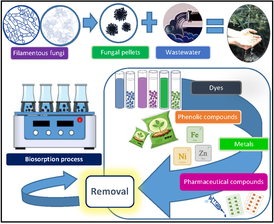
Table 1.
Biosorption of heavy metals by fungal pellets.
| Fungal Strain | Adsorbate | Operational Conditions | Adsorbate Concentration (mg·L−1) | Adsorption Capacity (mg·g−1) | Removal | Ref. |
| Aspergillus carbonarius | Cu(II) | pH: 5; agitation: 200 rpm; T: 25 °C; amount of pellets (L): 1 g (ww); time: 10 h | 100 | qexp: 1.7 | Not reported | [58] |
| Aspergillus flavus | Zn | pH: 5; agitation: 100 rpm; T: 34 °C; diameter of pellets: 1–3 mm; time: 6 days | 100 | Not reported | 40.9 ± 0.7% | [60] |
| Aspergillus fumigatus | 59.7 ± 0.5% | |||||
| Aspergillus lentulus | Cu(II) 1 | pH: 5; amount of pellets (L): 1.27–4.86 g·L−1 (dw) | 75–800 | qexp: 12.1–124.5 | 19.8–78.4% | [61] |
| pH: 2–8; amount of pellets (L): 1.71–4.89 g·L−1 (dw) | 80 | qexp: 1.7–15.2 | 3.6–79.8% | |||
| Cr(III) 1 | Amount of pellets (L): 4.03–4.64 g·L−1 (dw) | 1000–5000 | qexp: 171.0–331.5 | 26.0–79.4% | ||
| Ni(II) 1 | pH: 2–8; amount of pellets (L): 1.37–4.55 g·L−1 (dw) | 70–210 | qexp: 5.2–11.1 | 6.7–42.1% | ||
| 70 | qexp: 1.2–8.6 | 3.7–41.4% | ||||
| Pb(II) 1 | Amount of pellets (L): 0.46–4.67 g·L−1 (dw) | 500–4000 | qexp: 76.1–1120 | 12.9–71.0% | ||
| Aspergillus niger | Cu(II) | pH: 5.3; agitation: 100 rpm; T: 30 °C; amount of pellets (L): 1 g·L−1 (ww); time: 2 h | 30 | qmax: 8.1 | Not reported | [62] |
| Aspergillus niger 405 | Cu(II) | pH: 5; agitation: 200 rpm; T: 25 °C; amount of pellets: 1 g (ww); diameter of pellets: 1–3 mm; time: 10 h | 10 | qmax: 5.7 | Not reported | [53] |
| Zn(II) | qmax: 4.7 | |||||
| Ni(II) | qmax: 14.1 | |||||
| CrO42- | qmax: 7.2 | |||||
| Aspergillus japonicus | Fe(II) | pH: 2–10; agitation: 150 rpm; T: 30 °C; amount of pellets (L): 1 g (ww) | 25–100 | qmax: 1.3 | Not reported | [55] |
| Ni(II) | qmax: 1.2 | |||||
| Cr(VI) | qmax: 1.9 | |||||
| Fungal Strain | Adsorbate | Operational Conditions | Adsorbate Concentration (mg·L−1) | Adsorption Capacity (mg·g−1) | Removal | Ref. |
| Aspergillus japonicus | Hg(II) | qmax: 1.2 | Not reported | [55] | ||
| Industrial wastewater | pH: 2.1; agitation: 150 rpm; T: 30 °C; amount of pellets: 2.25 g (ww) | Ni(II): 44 Cr(VI): 90 | Ni(II): qmax: 1.16 Cr(VI): qmax: 2.57 | |||
| Funalia trogii | Cu(II) | pH: 5; agitation: 150 rpm; T: 30 °C; amount of pellets (L): 0.01 g·mL−1 (ww); diameter of pellets: 3–5 mm; time: 30 min | 10–300 | qmax: 23.89 | 61% | [63] |
| Lentinus edodes | Hg(II) Cd(II) Zn(II) | pH: 6; agitation: 400 rpm; T: 25 C, amount of pellets (L, A): 1 g·L−1 (ww); time: 2 h | 25–600 | (L): Hg(II) qmax: 358.1 Cd(II) qmax: 86.4 Zn(II) qmax: 37.7 | Not reported | [52] |
| (A): Hg(II) qmax: 419.1 Cd(II) qmax: 299.4 Zn(II) qmax: 63.3 | ||||||
| Penicillium sp. | Sr(II) Th(IV) U(VI) | pH: 5 for Sr(II) and U(VI); 3 for Th(IV); continuously stirred; T: 30 °C; amount of pellets (L, nFe3O4-pellets): 0.2 g·L−1; time: 48 h | Sr(II): 1–30 Th(IV): 1–130 U(VI): 1–130 | (L): Sr(II) qmax: 93.3 Th(II) qmax: 250.8 U(VI) qmax: 205.2 | Sr (II), pH 8: 100% Th(IV), pH 5: 100% U(VI), pH 7: 100% | [64] |
| Fe3O4-pellets: Sr(II) qmax: 109.9 Th(II) qmax: 280.8 U(VI) qmax: 223.9 | ||||||
| Penicillium chrysogenum | Cs | pH: 5; air lift column system; liquid to solid ratio (v/v): 10:1; diameter of pellets: 5 mm; time: 3 h | Cs: 13.3 | qmax: 119 | 50% | [65] |
| Sr | Sr: 8.8 | qmax: 92 | 39% | |||
| U | U: 23.8 | qmax: 147 | 62% | |||
| Fungal Strain | Adsorbate | Operational Conditions | Adsorbate Concentration (mg·L−1) | Adsorption Capacity (mg·g−1) | Removal | Ref. |
| Phanerochaete chrysosporium | As(III) | pH: 4; agitation: 150 rpm; ambient room T; amount of pellets (L): 0.25–1.5 g·L−1 (ww); time: 24 h | 0.2–1 | qmax: 5.5 | Not reported | [66] |
| Phanerochaete chrysosporium | Zn | pH: 4.5; agitation: 150 rpm; T: 30 °C; amount of pellets (L, nSe0-pellets): 3.2 g·L−1 (ww); time: 24 h | 10–50 | (L): qmax: 1.9–8.3 | (L): 56.2 ± 2.8% | [56] |
| (nSe0-pellets): qmax: 2.8–11.3 | (nSe0-pellets): 88.1 ± 5.3% | |||||
| Phanerochaete chrysosporium | Cu(II) | pH: 5; agitation: 150 rpm; T: 30 °C; amount of pellets: 0.01 g·mL−1 (ww); time: 30 min | 10–300 | qmax: 18.2 | 50% | [63] |
| Phanerochaete chrysosporium | Cd(II) | pH: 4.5; agitation: 150 rpm; T: 27 °C; amount of pellets (D): 0.1 g (dw); diameter of pellets: 1.58–2.03 mm; time: 18 h | 10–450 | qmax: 15.2 | Not reported | [67] |
| Pb(II) | 10–450 | qmax: 12.3 | ||||
| Binary system: Pb(II), Cd(II) | Cd(II): 10–450; Pb(II): 25–50 | Cd(II) qmax: 10–8.9 | ||||
| Pb(II): 10–450; Cd(II): 25–50 | Pb(II) qmax: 8.2–4.5 | |||||
| Phanerochaete chrysosporium | Pb(II) | pH: 5.5; agitation: 150 rpm; T: 27 °C; amount of pellets (L, Al. P): 2 g·L−1 (dw); diameter of pellets: 1.5–1.7 mm; time: 16 h | 50 | (L): qexp: 16.1 | 64.3% | [68] |
| (Al. P): qexp: 15.2–23.7 qmax: 144 | 60.6–94.7% | |||||
| Phanerochaete chrysosporium | Pb(II) | pH: 3–4; agitation: 200 rpm; T: 35 °C; amount of pellets (L, D): 90 mg (dw); time: 4 h | 5–50 | (L): qmax: 9 | Not reported | [69] |
| (D): qmax: 20 | ||||||
| pH: 5; agitation: 150 rpm; T: 25 °C; age of pellets (Ac. P, Al. P): 41, 168 h | 20 | (Ac. P): qmax: 12.8–13.8 | ||||
| (Al. P): qmax: 20.1–48.2 | ||||||
| Fungal Strain | Adsorbate | Operational Conditions | Adsorbate Concentration (mg·L−1) | Adsorption Capacity (mg·g−1) | Removal | Ref. |
| Phanerochaete chrysosporium | Cu(II) | pH: 6; agitation: 100 rpm; T: 25 °C; time: 4 h | 100 | qmax: 3.9 | Not reported | [70] |
| Phanerochaete chrysosporium | Cd(II) | pH: 6.2; T: 28 °C; amount of pellets: 0.2 g·mL−1 (ww); diameter of pellets: 0.2–2 cm; time: 2 h | 1124 | qexp: 84.5 | 34% | [71] |
| Pleurotus ostreatus | Cu(II) | Column experiments; pH: 3, 4; total column volume: 620 mL; flow rate: 1mL·min−1; amount of pellets (L): pH 3: 3.93 g (dw) pH 4: 3.37 g (dw) | 100 | pH: 3 qexp: 1.9 | Not reported | [58] |
| pH: 4 qexp: 7.9 | ||||||
| Rhizopus arrhizus | Ni(II) | pH: 8; agitation: 150 rpm; T: 35 °C; amount of pellets (L, D, Al. P): 3 g (ww); time: 72 h | 500 | (L): qmax: 315.6 | Not reported | [72] |
| (D): qmax: 125.4 | ||||||
| (Al. P): qmax: 357.6 | ||||||
| Rhizopus arrhizus | Cs | Air lift column system Liquid to solid ratio (v/v): 10:1 pH: 5; diameter of pellets: 5 mm; time: 3 h | Cs: 13.3 | qmax: 82 | 41% | [65] |
| Sr | Sr: 8.8 | qmax: 88 | >90% | |||
| U | U: 23.8 | qmax: 180 | 44% | |||
| Rhizopus nigricans | Pb(II) | Batch stirred tank Agitation: 300 rpm; amount of pellets: 25–200 g·L−1 (ww); diameter of pellets: 2.5 ± 0.5 mm | 20–300 | qmax: 83.5 | Not reported | [73] |
| Rhizopus nigricans | Li | pH: 5; agitation: 125 rpm; T: 22 ± 1 °C; diameter of pellets: 2.5 mm; time: 24 h | 10–1000 | qmax: 183.9 | Not reported | [74] |
| Al(III) | qmax: 163.0 | |||||
| Fe(II) | qmax: 466.4 | |||||
| Fe(III) | qmax: 407.7 | |||||
| Ni(II) | qmax: 201.2 | |||||
| Cu(II) | qmax: 360.4 | |||||
| Fungal Strain | Adsorbate | Operational Conditions | Adsorbate Concentration (mg·L−1) | Adsorption Capacity (mg·g−1) | Removal | Ref. |
| Rhizopus nigricans | Zn(II) | pH: 5; agitation: 125 rpm; T: 22 ± 1 °C; diameter of pellets: 2.5 mm; time: 24 h | 10–1000 | qmax: 235.1 | Not reported | [74] |
| Sr(II) | qmax: 278.0 | |||||
| Ag | qmax: 451.9 | |||||
| Cd(II) | qmax: 302.6 | |||||
| Pb(II) | qmax: 403.2 | |||||
| Rhizopus oryzae | Cu(II) | pH: 4; agitation: 150 rpm; T: 25–45 °C; amount of pellets (Al. P): 1 g (ww); diameter of pellets: 1–1.2 mm | 10–300 | qmax: 52.9–61.7 | [59] | |
| Trametes versicolor | Cd(II) | pH: 6.2; T: 28 °C; amount of pellets: 0.2 g·mL−1 (ww); diameter of pellets: 0.2–2 cm | 1124.1 | 109.5 | 43% | [71] |
1 Agitation: 180 rpm; T: 30 °C; diameter of pellets: 4–6 mm; time: 120 h. Pellets conditions: L: Live; A: Autoclaved; D: Dead; Ac. P: Acid pretreatment; Al. P: Alkali pretreatment; Fe3O4-pellets: pellets containing nanoparticles of Fe3O4; nSe0-pellets: pellets containing nanoparticles of Se. Weight of pellets: dw: dry weight; ww: wet weight. Adsorption capacity: qexp: experimental adsorption capacity (mg·g−1); qeq: adsorption capacity at the equilibrium (mg·g−1); qmax: maximum adsorption capacity obtained from Langmuir model (mg·g−1).
Table 2.
Summary of some pretreatments used in metal biosorption studies.
| Fungal Strain | Adsorbate | Pellet Pretreatment | Operational Conditions | Best Suited Pretreatment for Adsorption | Ref. |
|---|---|---|---|---|---|
| Lentinus edodes | Hg(II) Cd(II) Zn(II) | Thermal treatment | Pellets were heated at 90 °C for 15 min. | Thermal treatment | [52] |
| Penicillium sp. | Sr(II) Th(IV) U(VI) | Addition of nanoparticles of Fe3O4 | ~0.1 g of nano-Fe3O4 particles were added into a spore suspension cultivated at 140 rpm, 30 °C for 36 h. The mixed solution was incubated for further 72 h. | Fe3O4 addition | [64] |
| Phanerochaete chrysosporium | Zn | Addition of nanoparticles of Se (nSe0) | Pellets were grown with Na2SeO3 (10 mg Se L−1), 150 rpm, pH 4.5, and 30 °C for 96 h. | nSe0 addition | [56] |
| Phanerochaete chrysosporium | Cd(II) | Chemical and thermal inactivation | Pellets were inactivated by formaldehyde cross-linking and subsequent boiling in alkaline conditions for 45 min. | Not compared | [67] |
| Pb(II) | |||||
| Binary system: Pb(II), Cd(II) | |||||
| Phanerochaete chrysosporium | Pb(II) | Alkali pretreatment | Pellets were soaked in NaOH 0.1 M for 40 min. | Alkali pretreatment | [68] |
| Phanerochaete chrysosporium | Pb(II) | Autoclaving | Pellets were autoclaved (121 °C, 20 min). | Alkali pretreatment | [69] |
| Alkali pretreatment | Pellets were suspended in NaOH 0.1 M for 1 h. | ||||
| Acid pretreatment | Pellets were washed with HClO4 5 × 10⁻³ M for 5–40 min. | ||||
| Rhizopus arrhizus | Ni(II) | Alkali pretreatment | Pellets were treated separately with NaOH 0.1 M or NaCl 0.1 M for 30 min. Living (untreated) biomass was tested as a blank. | None (living biomass was best suited for Ni adsorption) | [72] |
| Rhizopus oryzae | Cu(II) | Thermal and alkali pretreatment | Pellets were boiled in NaOH 0.2 M (1:10 w/w) for 15 min. | Not compared | [59] |
Table 3.
Biosorption of dyes by fungal pellets.
| Fungal Strain | Adsorbate | Operational Conditions | Adsorbate Concentration (mg·L−1) | Adsorption Capacity (mg·g−1) | Removal | Ref. |
| Alternaria raphanin | Reactive Orange 107 | pH: 7; agitation: 150 rpm; T: 30 ± 2 °C; amount of pellets (L, A): 0.02 g·mL−1 (ww); time: 90, 105 min | 10–50 | (L): qexp: 8.8–21.5; qeq: 8.9–24.9 (A): qexp: 8.0–24.0; qeq: 10.8–27.7 | (L): ~47.9–80.4% (A): ~42.9–88.1% | [89] |
| Aspergillus foetidus | Reactive Black 5 | pH: 2.3; agitation 200 rpm; T: 30, 50 °C; amount of pellets (L, Al. P): 0.2 ± 0.05 g (ww) | 100 | (L): qmax: 65–76 | 67% | [90] |
| (Al. P): qmax: 92–106 | 97% | |||||
| Aspergillus lentulus | Acid Blue 120 | pH: 6.5; agitation 128 rpm; T: 30 °C; amount of pellets (L, A): 2 g·L−1 (dw) | 100 | (L): qmax: 101 | 91% | [91] |
| (A): qmax: 104.2 | ||||||
| pH: 6.5; agitation 180 rpm; T: 30 °C; amount of pellets (L): 0.75–3 g·L−1 (dw) | 100 | qexp: 33.9–58.2 | 38.7–98.3% | |||
| pH: 6.5; agitation 180 rpm; T: 30 °C; Amount of pellets (L): 2 g·L−1 | 100–900 | qexp: 54.6–210.7 | 46.9–96.3% | |||
| pH: 4–10; agitation 180 rpm; T: 30 °C; amount of pellets (L): 2 g·L−1 | 200 | qexp: 88.0–99.4 | 82.0–94.4% | |||
| pH: 6.5; agitation 180 rpm; T: 30–45 °C; amount of pellets (L): 2 g·L−1 | 200 | qexp: 94.7–99.4 | 90.0–95.6% | |||
| Aspergillus lentulus | Acid Blue 120 | pH: 6.5; agitation 180 rpm; T: 30 °C; amount of pellets (L): 2 g·L−1 (dw); time: 4, 5, 24 h | 100 | qmax: 181.8 | 96.7% | [92] |
| Acid Red 88 | qmax: 196.1 | 94.3% | ||||
| Pigment Orange 34 | qmax: 243.9 | 95.5% | ||||
| Fungal Strain | Adsorbate | Operational Conditions | Adsorbate Concentration (mg·L−1) | Adsorption Capacity (mg·g−1) | Removal | Ref. |
| Aspergillus lentulus | Basic Blue 9 | pH: 6.5; agitation 180 rpm; T: 30 °C; amount of pellets (L): 2 g·L−1 (dw); time: 4, 5, 24 h | 30 | qmax: 30.7 | 21.8–35.4% | [92] |
| Basic Violet 10 | qmax: 13.9 | 54.9–90.9% | ||||
| Acid Navy Blue and Fast Red A | 100 | Not reported | 94.2% | |||
| Acid Navy Blue and Methylene Blue | 50 | Not reported | 70.1% | |||
| Aspergillus lentulus | Acid Violet 19 | pH: 6.5; agitation 180 rpm; T: 30 °C; inoculum: 5% spore suspension of 6.25·106 mL; time: 20 h, 50 h | 100 | Not reported | 49–70% | [93] |
| Acid Blue 120 | 99% | |||||
| Acid Blue 89 | 92–98% | |||||
| Acid Red 88 | 98–99% | |||||
| Pigment Orange 34 | 91–99% | |||||
| Acid Blue 120 | 100–900 | qexp: ~25–212 | 100% for all the tested concentrations | |||
| Aspergillus niger ZJUBE-1 | Congo Red | pH: 2–5; agitation 120 rpm; T: 28 °C amount of pellets (L): 4 g·L−1 (ww) diameter of pellets: 2–4 mm; time: 12 h | 25–300 | qexp: 14.6–152.0 qmax: 263.2 | 99% | [2] |
| Acid Blue 29 Acid Red 27 Amido Black 10 B Congo Red Eriochrome Black T Orange II Methyl Blue Naphthol Green | pH: 6; agitation 120 rpm; T: 28 °C; amount of pellets (L): 2 g·L−1(ww); diameter of pellets: 2–4 mm; time: 24 h | 50 | Not reported | Orange II: 77.6% All the other tested dyes: >94% | ||
| Fungal Strain | Adsorbate | Operational Conditions | Adsorbate Concentration (mg·L−1) | Adsorption Capacity (mg·g−1) | Removal | Ref. |
| Aspergillus niger ZJUBE-1 | Eriochrome Black T | Batch Reactor pH: 6; aeration: 1 L·min−1; T: 25 °C; amount of pellets (L): 30 g (ww); diameter of pellets: 2–4 mm; time: 12 h | 50 | qmax: 322.6 | >90% | [2] |
| Aspergillus niger | Procion Red MX-5B | pH: 4; T: 30 ± 1 °C; time: 3 h | 0.2 | Not reported | ~30% | [94] |
| Aspergillus oryzae | Procion Violet Red H3R | pH: 2.5; amount of pellets (L, A): 3.7·10−4–4.5−3 g·mL−1; Time: 120 min | 62.5 | (L): qexp: 9.6–25.0 (A): qexp: 11.0–26.3 | Not reported | [95] |
| Procion Red HE7B | pH: 2.5; amount of pellets (L, A): 3.7−4–0.03 g·mL−1 Time: 120 min | 300 | (L): qexp: 9.6–20.2 (A): qexp: 9.6–32.4 | |||
| Funalia trogii | Astrazon Black | Repeated batch system: 5 cycles agitation 150 rpm; T: 30 °C; pellets (L, D); time: 24 h | 100 | Not reported | (L): 1rst–5th use: ~92–40% (D): 1rst–3rd use: ~76–40% | [88] |
| Penicillium chrysogenum MT-6 | Reactive Black 5 | pH: 5; agitation 150 rpm; T: 28 °C amount of pellets: 3.83 g·L−1 (ww) time: 100 h | 300 | Not reported | 89% | [96] |
| Penicillium chrysosporium | Astrazon Black | Repeated batch system: 5 cycles agitation 150 rpm; T: 30 °C pellets (L, D); time: 24 h | 100 | - | (L): 1rst–5th use: ~92–28% (D): 1rst–4th use: ~72–10% | [88] |
| Penicillium oxalicum | Reactive Blue 19 | pH: 2; agitation 150 rpm; T: 20 °C amount of pellets (L): 0.25 g (ww) diameter of pellets: 2 mm; time: 24 h | 100 | qmax: 160 | Reactive Blue 19: 60–91% | [97] |
| Reactive Red 241 | qmax: 122 | |||||
| Reactive Yellow 145 | qmax: 137 | |||||
| Fungal Strain | Adsorbate | Operational Conditions | Adsorbate Concentration (mg·L−1) | Adsorption Capacity (mg·g−1) | Removal | Ref. |
| Penicillium oxalicum | Cu-Complex Reactive Dye Blue 21 | pH: 7; agitation 120 rpm; T: 30 °C; amount of pellets (L): 119 ± 3.9–129 ± 4.1 mg (dw); diameter of pellets: 3.83 mm; time: 48 h | 100–300 | qmax: 77.5 ± 3.2–336 ± 6.8 | 99.7–100% | [98] |
| Penicillium sp. | Acid Violet | pH: 5.7; agitation 150 rpm; T: 35 °C amount of pellets (L): 0.01 g·mL−1 (ww); time: 12 h | 30 | qmax: 5.9 | Not reported | [84] |
| Phanerochaete chrysosporium | Indigo dye and dyeing wastewater | Repeated batch system: 5 cycles; T: 30 °C; amount of pellets (L, D): 0.0092 g·mL−1 | Not reported | Not reported | (L): 1rst–5th use: 94.9–88.3% (D): 1rst–5th use: 36.5–38.7% | [99] |
| Trametes versicolor | Astrazon Black | Repeated batch system: 5 cycles; agitation 150 rpm; T: 30 °C; pellets (L, D); time: 24 h | 100 | Not reported | (L): 1rst–5th use: ~92–20% (D): 1rst–4th use: ~62–28% | [88] |
| Trametes versicolor | Congo Red | Bubble reactor type; pH: 7; aeration: 100 cm·s−1; T: 30 °C inoculum: 106 spores·mL−1; amount of pellets (L, A, Ac. P, Al. P): 1 g; time: 90 min | 10–50 | (L): qexp: 10–42.4; qmax: 50.8 | Not reported | [100] |
| (A): qexp: 10–46.1; qmax: 51.8 | ||||||
| (Ac. P): qexp: 6.9–26.2; qmax: 42.2 | ||||||
| (Al. P): qexp: 6.1–22.4; qmax: 38.2 | ||||||
| Trametes versicolor | Congo Red | Rotating biological contactor T: 30 °C; inoculum: 1 mL fungal spore suspension (106 spores); pellets (L, A); time: 65 min | 10–50 | (L): qmax: 38.9 (A): qexp: 10.0–36.5; qmax: 41.49 | Not reported | [100] |
| Fungal Strain | Adsorbate | Operational Conditions | Adsorbate Concentration (mg·L−1) | Adsorption Capacity (mg·g−1) | Removal | Ref. |
| Trametes versicolor | Congo Red | pH: 2–10; agitation 150 rpm; T: 27 ± 2 °C; amount of pellets (L, A, Ac. P, Al. P): 1 g/50 mL (ww); time: 90 min | 10–50 | (L): qexp: 10–44; qmax: 52.9 | 87.9–100% | [78] |
| (A): qexp: 10–47.7 | 95.4–100% | |||||
| (Ac. P): qmax: 44.1 | 48.0–66.7% | |||||
| (Al. P): qmax: 39.5 | 55.5–74.1% | |||||
| Trichoderma harzianum | Acid Brilliant Red B | pH: 3; agitation: 120 rpm; T: 30 °C; inoculum: 5% v/v spore suspension (A = 0.5/cm at 600 nm) pellets (G, L, D); time: 72 h | 100–400 | (G): qmax: 86.5 ± 7.4–365 ± 25.2 (L): qmax: 73.0 ± 2.9–183 ± 12.6 (D): qmax: 65.7 ± 7.1–136 ± 14.1 | Not reported | [101] |
| Trichoderma harzianum | Rhodamine 6G | pH: 8; agitation: 125 rpm; T: 30 °C; amount of pellets (A): 0.02 g·mL−1 (ww); time: 90 min | 10–50 | qexp: 0.4–1.9 qmax: 3.5 | 87–77% | [13] |
| Erioglaucine | pH: 4; agitation: 125 rpm; T: 30 °C; amount of pellets (A): 0.02 g·mL−1 (ww); time: 75 min | qexp: 0.4–1.9 qmax: 3.14 | 88–76% |
Pellets conditions: L: live; A: autoclaved; D: dead; Ac. P: acid pretreatment; Al. P: alkali pretreatment, G: growing. Weight of pellets: dw: dry weight, ww: wet weight. Adsorption capacity: qexp: experimental adsorption capacity (mg·g−1); qeq: adsorption capacity at the equilibrium (mg·g−1); qmax: maximum adsorption capacity obtained from Langmuir model (mg·g−1).
Table 4.
Biosorption of phenolic compounds by fungal pellets.
| Fungal Strain | Adsorbate | Operational Conditions | Adsorbate Concentration (mg·L−1) | Adsorption Capacity (mg·g−1) | Removal | Ref. |
|---|---|---|---|---|---|---|
| Anthracophyllum discolor | Pentachlorophenol | pH: 5–5.5; agitation: 120 rpm T: 25 °C; amount of pellets (L): 20 ± 0.2 mg (dw); time: 24 h | 5–10 | Not reported | >80% | [111] |
| Funalia trogii | Phenol | pH: 2–11; agitation: 100 rpm; T: 25 °C; amount of pellets (L, A): 0.25–2 g·L−1; time: 6 h | 200 | (L): qmax: 132.6 (A): qmax: 132.6 | Not reported | [110] |
| 2-Chlorophenol | (L): qmax: 132.6 (A): qmax: 132.6 | |||||
| Penicillium oxalicum | Phenol | pH: 6; agitation: 150 rpm; T: 30 °C; amount of pellets (A): 0.25 g (ww); time: 24 h | 10–100 | qmax: 0.01 | Not reported | [9] |
| p-Cresol | qmax: 0.02 | |||||
| Naphthol | qmax: 0.06 | |||||
| p-Toluidine | qmax: 0.03 | |||||
| 1-Naphthalenamine | qmax: 0.15 | |||||
| p-Toluenesulfonic acid | qmax: 0.01 | |||||
| Phanerochaete chrysosporium | Phenol | pH: 5.5; agitation: 4000 rpm T: 22.5 °C; amount of pellets (L): 0.3 g·L−1; time: 2 h | 25–100 | qmax: 2.8–13.5 qeq: 6.7 | Not reported | [114] |
| Phanerochaete chrysosporium | Phenol | pH: 5; agitation: 150 rpm; T: 22.5 ± 2.5 °C; amount of pellets (A): 5 g·L−1; time: 3 h | 12.5–50 | qmax: 0.09–0.19 qeq: 0.09–0.19 | Not reported | [115] |
| Trametes versicolor | Pentachlorophenol | pH: not reported; agitation: 70 rpm; T: 25 °C; amount of pellets (D): 0.4 g·L−1; time: 24 h | 0.05 | Not reported | >60% | [116] |
| 4-Tert-butylphenol | 10 ± 14% | |||||
| Bisphenol A | 26 ± 5% | |||||
| 4-Tert-octophenol | >60% | |||||
| Benzophenone | 10 ± 2% | |||||
| Octocrylene | 27 ± 2% |
Pellets conditions: L: live; D: dead. Weight of pellets: dw: dry weight; ww: wet weight. Adsorption capacity: qexp: experimental adsorption capacity (mg·g−1); qeq: adsorption capacity at the equilibrium (mg·g−1); qmax: maximum adsorption capacity obtained from Langmuir model (mg·g−1).
Table 5.
Biosorption of other organic compounds by fungal pellets.
| Fungal Strain | Adsorbate | Operational Conditions | Adsorbate Concentration (mg·L−1) | Adsorption Capacity (mg·g−1) | Removal | Ref. |
|---|---|---|---|---|---|---|
| Aspergillus fumigatus | Humic acid | pH: 4; agitation: 150 rpm; T: 30 °C; age culture: 2–10 days; amount of pellets (L): 0.5 g (ww); time: 1 h | 24 ± 0.5 | 2 days-grown pellets: qexp: 6.8 | Not reported | [126] |
| 10 days-grown pellets: qexp: 9.0 | ||||||
| Ganoderma lucidum | Picloram | pH: 3.76; agitation: 150 rpm; T: 25 °C; amount of pellets (L, A): 0.05 g (dw); time: 2 h | 2415 | (L): qeq: 6.6 (A): qeq: 7.9 | Not reported | [122] |
| Trametes sp. | (L): qeq: 5.6 (A): qeq: 6.0 | |||||
| Trametes versicolor | Acid clofibric | pH: not reported; agitation: 70 rpm; T: 25 °C; amount of pellets (D): 0.4 g·L−1; time: 24 h | 0.05 | Not reported | 15 ± 3% | [116] |
| Atrazine | 9 ± 1% | |||||
| Ametryn | 4 ± 8% |
Pellets conditions: L: live; A: autoclaved; D: dead. Weight of pellets: dw: dry weight; ww: wet weight. Adsorption capacity: qexp: experimental adsorption capacity (mg·g−1); qeq: adsorption capacity at the equilibrium (mg·g−1).
Table 6.
Biosorption of pharmaceutical compounds by fungal pellets.
| Fungal Strain | Adsorbate | Operational Conditions | Adsorbate Concentration (mg·L−1) | Removal | Ref. | ||
| Ganoderma lucidum | Carbamazepine | Agitation: 135 rpm; T: 25 °C; time: 6 days; under dark conditions; amount of pellets (A): 0.5 ± 0.1 g (dw) | 0.05–0.18 | 13 ± 1% | [137] | ||
| Diclofenac | 34 ± 3% | ||||||
| Iopromide | 18 ± 1% | ||||||
| Venlafaxine | 11 ± 1% | ||||||
| Ganoderma lucidum | Ibuprofen | Agitation: 135 rpm; T: 25 °C; age of culture: 7 days; amount of pellets (A): 2 g·L−1 (ww) | 10 | ~50% | [135] | ||
| Carbamazepine | ~10% | ||||||
| Clofibric acid | ~23% | ||||||
| Irpex lacteous | Carbamazepine | Agitation: 135 rpm; T: 25 °C; time: 6 days; under dark conditions; amount of pellets (A): 0.5 ± 0.1 g (dw) | 0.05–0.18 | 5 ± 1% | [137] | ||
| Diclofenac | 26 ± 3% | ||||||
| Iopromide | 5 ± 1% | ||||||
| Venlafaxine | 6 ± 1% | ||||||
| Irpex lacteous | Ibuprofen | Agitation: 135 rpm; T: 25 °C; age of culture: 7 days; amount of pellets (A): 2 g·L−1 (ww) | 10 | Not removed | [135] | ||
| Carbamazepine | ~20% | ||||||
| Clofibric acid | ~5% | ||||||
| Phanerochaete chrysosporium | Ibuprofen | Agitation: 135 rpm; T: 25 °C; age of culture: 7 days on malt extract medium; amount of pellets (A): 2 g·L−1 (ww) | 10 | Not removed | [135] | ||
| Carbamazepine | ~31% | ||||||
| Clofibric acid | ~26% | ||||||
| Phanerochaete chrysosporium | Ibuprofen | Agitation: 135 rpm; T: 25 °C; age of culture: 7 days on malt extract medium; amount of pellets (A): 2 g·L−1 (ww) | 10 | Not removed | [135] | ||
| Carbamazepine | ~8.5% | ||||||
| Clofibric acid | ~27% | ||||||
| Trametes hirsuta | 0.0001 | L: | A: | B: | [138] | ||
| Carbamazapine | ~36% | ~28% | - | ||||
| Acid clofibric | ~5% | 100% | ~60% | ||||
| Naproxen | 100% | ~90% | - | ||||
| Ketoprofen | 100% | ~12% | ~14% | ||||
| Ibuprofen | 100% | ~90% | ~75% | ||||
| Fungal Strain | Adsorbate | Operational Conditions | Adsorbate Concentration (mg·L−1) | Removal | Ref. | ||
| L: | A: | B: | |||||
| Trametes hirsuta | Indomethacin | 0.0001 | 100% | 100% | ~10% | [138] | |
| Acetaminophen | ~76% | ~30% | ~45% | ||||
| Fenofibrate | 100% | 100% | 100% | ||||
| Amoxicillin | 100% | ~30% | ~38% | ||||
| Ioperamide | ~24% | ~10% | ~10% | ||||
| Trametes versicolor | Carbamazepine | Agitation: 135 rpm; T: 25 °C; time: 6 days; under dark conditions; amount of pellets (A): 0.5 ± 0.1 g (dw) | 0.05–0.18 | 8 ± 1% | [137] | ||
| Diclofenac | 42 ± 12% | ||||||
| Iopromide | 8 ± 1% | ||||||
| Venlafaxine | 6 ± 1% | ||||||
| Trametes versicolor | Ibuprofen | Amount of pellets: 0.4 g·L−1 (ww) | 0.05 | 32 ± 1% | [116] | ||
| Gemfibrozil | >60% | ||||||
| Diclofenac | >60% | ||||||
| Triclosan | >60% | ||||||
| Ketoprofen | 10 ± 15% | ||||||
| Naproxen | 14 ± 5% | ||||||
| Carbamazepine | 6 ± 5% | ||||||
| Primidone | 24 ± 34% | ||||||
| Amitriptyline | 8 ± 8% | ||||||
| Trametes versicolor | Estriol | Amount of pellets: 0.4 g·L−1 (ww) | 0.05 | 13 ± 4% | [116] | ||
| Estrone | >60% | ||||||
| 17α-Ethinylestradiol | >60% | ||||||
| 17β-Estradiol | >60% | ||||||
| 17β-Estradiol-17acetate | >60% | ||||||
| Fungal Strain | Adsorbate | Operational Conditions | Adsorbate Concentration (mg·L−1) | Removal | Ref. | ||
| Trametes versicolor | Enterolactone | Amount of pellets: 0.4 g·L−1 (ww) | 0.05 | 3 ± 15% | [116] | ||
| Formononetin | 15 ± 5% | ||||||
| Trametes versicolor | Ibuprofen | Agitation: 135 rpm; T: 25 °C; amount of pellets: 2 g·L−1 (ww) | 10 | ~25% | [116] | ||
| Carbamazepine | ~10% | ||||||
| Clofibric acid | ~5% | ||||||
Pellets conditions: L: live; A: autoclaved; B: Biocide pretreatment. Weight of pellets: dw: dry weight; ww: wet weight.
© 2020 by the authors. Licensee MDPI, Basel, Switzerland. This article is an open access article distributed under the terms and conditions of the Creative Commons Attribution (CC BY) license (http://creativecommons.org/licenses/by/4.0/).
Share and Cite
MDPI and ACS Style
Legorreta-Castañeda, A.J.; Lucho-Constantino, C.A.; Beltrán-Hernández, R.I.; Coronel-Olivares, C.; Vázquez-Rodríguez, G.A. Biosorption of Water Pollutants by Fungal Pellets. Water 2020, 12, 1155. https://doi.org/10.3390/w12041155
AMA Style
Legorreta-Castañeda AJ, Lucho-Constantino CA, Beltrán-Hernández RI, Coronel-Olivares C, Vázquez-Rodríguez GA. Biosorption of Water Pollutants by Fungal Pellets. Water. 2020; 12(4):1155. https://doi.org/10.3390/w12041155
Chicago/Turabian StyleLegorreta-Castañeda, Adriana Jazmín, Carlos Alexander Lucho-Constantino, Rosa Icela Beltrán-Hernández, Claudia Coronel-Olivares, and Gabriela A. Vázquez-Rodríguez. 2020. "Biosorption of Water Pollutants by Fungal Pellets" Water 12, no. 4: 1155. https://doi.org/10.3390/w12041155
Note that from the first issue of 2016, this journal uses article numbers instead of page numbers. See further details here.