The Influence of Residual Coagulant Al on the Biofilm EPS and Membrane Fouling Potential in Wastewater Reclamation

1
School of Environment, Northeast Normal University, Changchun 130117, China
2
Jilin Engineering Lab for Water Pollution Control and Resources Recovery, Northeast Normal University, Changchun 130117, China
*
Author to whom correspondence should be addressed.
Water 2020, 12(4), 1056; https://doi.org/10.3390/w12041056
Submission received: 23 February 2020
/
Revised: 24 March 2020
/
Accepted: 27 March 2020
/
Published: 8 April 2020
(This article belongs to the Section Wastewater Treatment and Reuse)
Abstract
:Biofouling is inevitable in wastewater reclamation when using membrane technology. In particular, the extracellular polymeric substances (EPS) from biofilm is a major contributor to biofouling. Coagulation is critical in the process of reusing wastewater before membrane treatment, and residual coagulants (e.g., Al salts) are able to alter the characteristics of the biofilm EPS. However, the distribution of residual Al across varying biofilm EPS fractions and its effect on the membrane fouling potential resulting from biofilm EPS remains unclear. We found that 34% of the residual Al was present in the soluble EPS (S-EPS), 26% in the loosely bound EPS (LB-EPS) and 40% in the tightly bound EPS (TB-EPS). Moreover, compared with the control groups, the residual Al in biofilm induced more biofilm formation and more EPS formation. Al reduced the zeta potential and increased the hydrophobicity of the EPS. These changes induced a significant rise in the membrane fouling potential of S-EPS and LB-EPS. This work provides coagulation support for wastewater reclamation using membrane technology.
1. Introduction
Increases in the demand of high-quality water have generated advances in wastewater treatment. In particular, membrane technology has been developed for the process of reusing wastewater. It is easy to operate, stable and its effluent is of high quality [1]. However, membrane fouling is a critical limitation of this technology, seriously reducing fluxes and affecting effluent quality [2]. Membrane fouling can be grouped into four categories: the deposition of particles, inorganic pollution, organic pollution and biofouling [3]. In particular, biofouling is the most difficult to control and is predominantly caused by the biofilm [4]. More specifically, previous research has indicated that biofouling is the result of extracellular polymeric substances (EPS) surrounding the bacteria within the biofilm.
A biofilm is an accumulation of bacteria attached to a solid surface and is generally composed of microbial cells, EPS and inorganic materials [5]. EPS is a gel-like matrix that binds cells, accounting for 80% of the biofilm [6]. EPS contains high molecular-weight biopolymers originating from bacterial secretion and adsorbed organic matter [7]. EPS can be grouped into three categories: soluble EPS (S-EPS), loosely bound EPS (LB-EPS) and tightly bound EPS (TB-EPS). Different EPS fraction characteristics may be affected by environmental factors reported by Desmond [8] and different EPS fraction matrix compositions have different fouling potential to the membrane.
Chemical coagulation plays a key role in wastewater reclamation. Aluminum salt coagulants are commonly used in wastewater reclamation on account of their high efficiency and low cost [9]. However, aluminum ions remain in the effluent, with observed concentrations within the range of 0.54–2.12 mg/L [10]. In previous work, Cui (2016) investigated the role which aluminum plays in wastewater reclaiming [10]. In another work, Cui (2018) reported that residual Al could stimulate biofilm formation, thus changing the EPS characteristics when combined with proteins [11]. However, the effects of aluminum on the different EPS fractions (S-EPS, LB-EPS and TB-EPS) and its influence on the post membrane treatment still remain unclear.
In summary, wastewater reclamation has become a common phenomenon due to the lack of water sources, yet it is limited by biofouling. However, the influence of residual coagulant on different biofilm EPS fractions as well as the post biofouling mechanism is not well known. In this study, we investigated the roles of residual aluminum in different fractions of EPS by identifying the Al amounts present in the fractions and the subsequent changes of each fraction. Particular attention was placed on the fouling potential of the changed EPS fractions under an ultrafiltration system. We aim to gain a comprehensive insight into the spatial distribution of the aluminum present in biofilm as well as a deep understanding of the effects of residual coagulant aluminum on the physicochemical characteristics of each EPS fraction.
2. Materials and Methods
2.1. Al Concentration Selection and Biofilm Cultivation
Biofilm formation was investigated at Al concentration of 0.5 mg–Al/L, which is a typical residual Al concentration [11]. Mixed bacteria from activated sludge (South Wastewater Treatment Plant, Changchun, China) were selected as the model bacteria. Biofilms were cultivated using synthetic wastewater composed of glucose 0.094 g/L, NH4Cl 0.064 g/L, K2HPO4·3H2O 0.007 g/L, KH2PO4 0.004 g/L, MgSO4 0.002 g/L, CaCl2 0.005 g/L [12], and 1 mL/L trace element solution (FeSO4·7H2O 1.36 g/L, Na2MoO4·2H2O 0.24 g/L, CuSO4·5H2O 0.25 g/L, ZnSO5·7H2O 0.58 g/L, NiCl2·6H2O 0.10 g/L, MnSO4·H2O 1.01 g/L [13]). The biofilm was characterized after 35 days of cultivation.
2.2. Reactor Operation
Two self-designed reactors (308 × 184 × 350 mm cuboids) were applied with a continuous water flow for the development of biofilms. The influent for one reactor contained aluminum, while the other, denoted as the control, did not contain aluminum. The inlet is on the right-hand bottom of the reactor, and the outlet is on the left-hand top of the reactor. The synthetic reclaimed water was pumped through the inlet (15 mL/min), and the hydraulic retention time is 17 h. Four glass carriers (84 × 62 × 250 mm) were placed in the reactor, with the potential to hold 30 pieces of glass for biofilm cultivation. Dissolved oxygen was controlled within 6.25–7.17 mg/L, the pH within 7.38–7.58 and the temperature maintained at 25 °C. The reactors were operated for 35 days.
2.3. EPS Extraction
The S-EPS, LB-EPS and TB-EPS fractions were extracted by a combination of centrifugation and thermal extraction [14]. More specifically, approximately 200 mL of fresh biofilm collected from glass carriers was placed in 500 mL centrifuge tubes and centrifuged at 3000× g for 10 min. The supernatant was regarded as the S-EPS fraction. The residues were resuspended to the initial volume in 0.05% (w/w) NaCl solution, and subsequently sonicated at 20 kHz for 2 min, the liquid was centrifuged at 8000× g for 10 min. The supernatant was deemed the LB-EPS fraction. The residues were resuspended at their initial volume, following sonicated at 20 kHz for 2 min and heated at 60 °C for 30 min. The liquid was centrifuged at 12,000× g for 20 min, and the collected supernatant was the TB-EPS. Finally, the EPS fractions were filtered using a 0.45 μm filter membrane before testing.
2.4. EPS Fraction Membrane Fouling Potential
The amount of total EPS was measured using dissolved organic carbon (DOC) concentrations of 0.1 g/L for the different EPS fractions that were prepared. The membrane fouling potential of the EPS fractions was determined directly in terms of normalized flux using a laboratory-scale ultrafiltration cup (400 mL volume, 35 mm effective diameter). The pressure of 0.15 MPa was applied utilizing nitrogen gas. The permeated flux (J) was recorded every 3 min, with the first recording defined as the initial permeate (J0). The normalized flux was then determined as J/J0.
2.5. Analytical Methods
2.5.1. Biofilm Dry Weight
Samples were dried at 105 °C in oven, and the dry weight the biofilm per unit area was measured.
2.5.2. Proteins and Polysaccharides Concentrations
The proteins content present in EPS was measured using a Folin’s phenol reagent, with bovine serum albumin as the standard. Polysaccharide amounts in the EPS were determined using the anthrone·H2SO4 method with glucose as the standard [15].
2.5.3. Spectrum Analysis
The EPS from all extracted samples was frozen and dried at −60 °C, with compositions compared using a Fourier-transform infrared spectrophotometer (FTIR) (CARY 630, Agilent Technologies Inc., Santa Clara, CA, USA).
Fluorescent compounds were distinguished from complex EPS mixtures using three-dimension excitation-emission matrix (3D-EEM) fluorescence spectroscopy. More specifically, all EPS fractions were prepared to 0.1 g/L in deionized water and their 3D-EEM spectra were measured using a fluorescence spectrophotometer (LS 55, Perkin Elmer, Waltham, MA, USA). The emission and excitation spectra ranged over 200–550 nm at 10 nm sampling interval. The scanning speed was held at 1200 nm/min.
2.5.4. Hydrophilicity and Hydrophobicity
In order to evaluate the EPS hydrophilicity and hydrophobicity cultivated by different concentrations of Al, the contact angle between the EPS and a glass sheet was characterized using a Drop Shape Analyzer (DSA25, KRUSS, Suzhou, China).
2.5.5. Zeta Potential
The Zeta potential of each EPS fraction was tested using Zetasizer Nano-ZS (Malvern Instruments, Malvern, UK). S-EPS, LB-EPS and TB-EPS concentrations were adjusted to 1 g/L (in deionized water). All tests were performed in triplicate.
The flocculating efficiencies of EPS were acquired using kaolin suspension [16]. Briefly, 0.1 mL of EPS (1 g/L), 4.65 mL kaolin suspension (5 g/L) and 0.25 mL of CaCl2 (90 mM) were mixed. The substances in the tube were mixed using a vortex machine at 1300 rpm for 30 s, and then left to sit for 5 min. The absorbance of sample (Abs550) and control (Abs550,b) were tested at 550 nm wavelength utilizing a UV-Vis spectrophotometer (UV-2800A, Unico, Shanghai, China). All tests were performed in triplicate.
Flocculating efficiency % = (Abs550,b−Abs550)/Abs550,b
2.6. Aluminum Distribution Measurements
The aluminum in different EPS fractions was determined by the EPS acidic digestion method [5]. Briefly, 2 mL nitric acid (69%) and 1 mL hydrogen peroxide (30%) were added to 3 mL EPS. The mixture was then heated for 2 h at 120 °C. After this, the sample was placed in a 10 mL capacity bottle, and subsequently passed through a 0.22 μm filter membrane. Aluminum concentrations were then determined by inductive coupled plasma (ICP-AES).
3. Results and Discussion
3.1. Acceleration of Biofilm EPS Fractions Formation via Residual Al
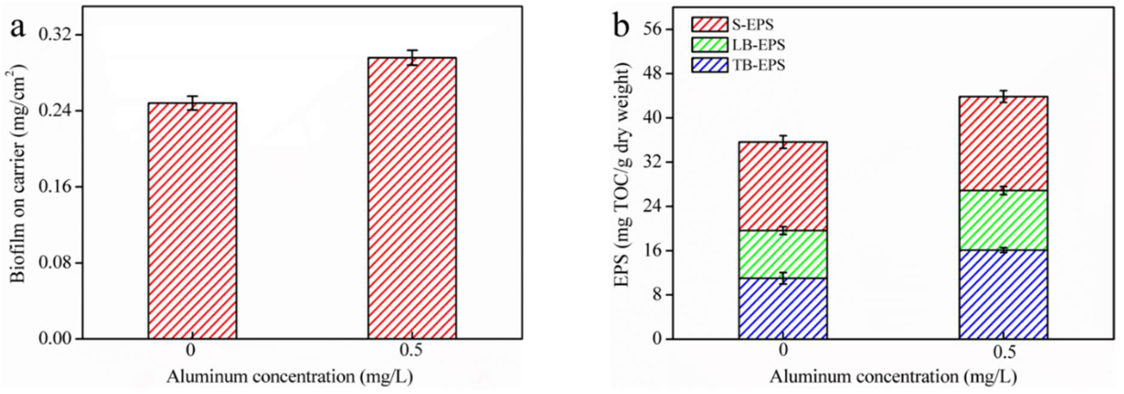
For the aluminum concentration of 0 mg/L, the dry weight of biofilm was 0.25 ± 0.01 mg/cm2. The dry weight increased as 0.29 ± 0.01 mg/cm2 with the aluminum concentration of 0.5 mg/L (p < 0.01) (Figure 1a). This suggests that the addition of aluminum significantly impacted the formation of the biofilm. This may be explained by the promotion of the EPS secretion attributed to aluminum ions, resulting in tight bonding with bacteria and an increase in biofilm weight [11]. Aluminum ions may neutralize the negatively charged EPS groups, reducing the electrostatic repulsion between cells while enhancing affinity, thus facilitating biofilm growth [17]. Moreover, the pH of the reaction system was approximately 7.5, while the majority of aluminum ion in water was in the form of hydroxides, which can adsorb more nutrients, benefiting microbial growth [18].
Aluminum ions were observed to enhance the production of the EPS fractions (Figure 1b). More specifically, TB-EPS content increased sharply from 11 to 16.1 mg TOC/g dry weight (DW), LB-EPS increased from 8.62 to 10.77 mg TOC/g DW and S-EPS increased slightly from 16.01 to 16.99 mg TOC/g DW. This indicates that the aluminum caused different degrees of variation within different biofilm EPS fractions, which may be attributed to the different aluminum distributions in the biofilm. In particular, the increased content was observed to be higher for TB-EPS and LB-EPS compared to that of S-EPS. This can be explained by the thick barrier resulting from the bound EPS for cells in adverse atmospheres (e.g., in the presence of aluminum ion) [19].
3.2. Al Distribution and Changes in EPS Fractions
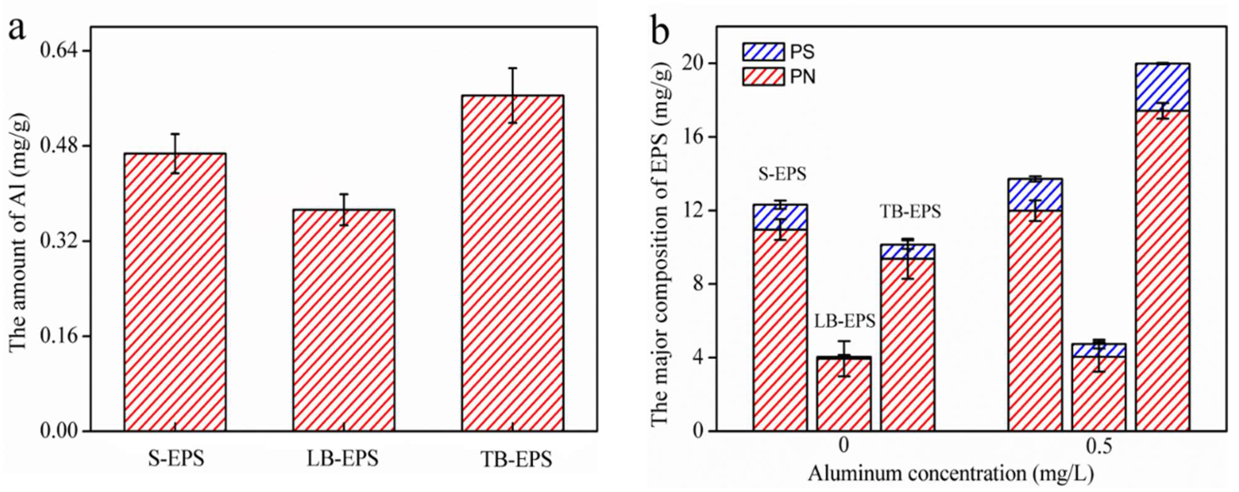
The aluminum concentrations determined in S-EPS, LB-EPS and TB-EPS fractions were 0.47 mg/g, 0.37 mg/g and 0.56 mg/g, respectively (Figure 2a). Biofilms are composed of cellular aggregates and EPS. These components can react with dissolved species by chemical or physical interactions, with such reactions determined by the presence of binding sites [5]. S-EPS is able to move freely between the biofilm and liquor. Meanwhile, LB-EPS, the outer layer of bound EPS, is a loose slime layer without an obvious edge, and TB-EPS exhibits a certain morphology. The different physicochemical properties of each EPS fraction (e.g., flocculation potential, functional groups, binding sites and EPS depth) were distinct, thus leading to variations in the aluminum distribution between fractions [6].
The S-EPS, LB-EPS and TB-EPS contents were 16.1 mg TOC/g DW, 10.77 mg TOC/g DW and 16.99 mg TOC/g DW, respectively. As the EPS concentration increased, a greater amount of aluminum was combined. The increased aluminum may have led to a greater amount of secreted EPS by the bacteria in order to protect the cell [20]. Total protein and polysaccharide contents, the two major EPS components, were in proportion to the corresponding EPS fraction amounts. Polysaccharide amounts varied from 1.35 to 1.73 mg/g, 0.1 to 0.7 mg/g and 0.7 to 2.56 mg/g in the S-EPS, LB-EPS and TB-EPS fractions, respectively. Protein amounts varied from 10.96 to 11.99 mg/g, 3.94 to 4.04 mg/g and 9.37 to 17.42 mg/g in the S-EPS, LB-EPS and TB-EPS fractions, respectively (Figure 2b). With the aluminum, polysaccharide amounts increased by 28%, 579% and 233% in S-EPS, LB-EPS and TB-EPS, respectively. Protein amounts increased by 9%, 3% and 85% in S-EPS, LB-EPS and TB-EPS, respectively. The strong viscosity of polysaccharides is responsible for the adhesion interaction in cells, and the polysaccharides are also more capable of accumulating on the membrane compared with proteins. Proteins played a vital role during the initial biofilm formation [21]. They are a key factor in the stabilization of the EPS structure [22], which was dominant within the bottom biofilm layer, thus accelerating bacteria attachment. The bacterial cells responded to the exogenous stimulation, thus secreting more proteins and polysaccharides in order to resist cell damage from aluminum [23].
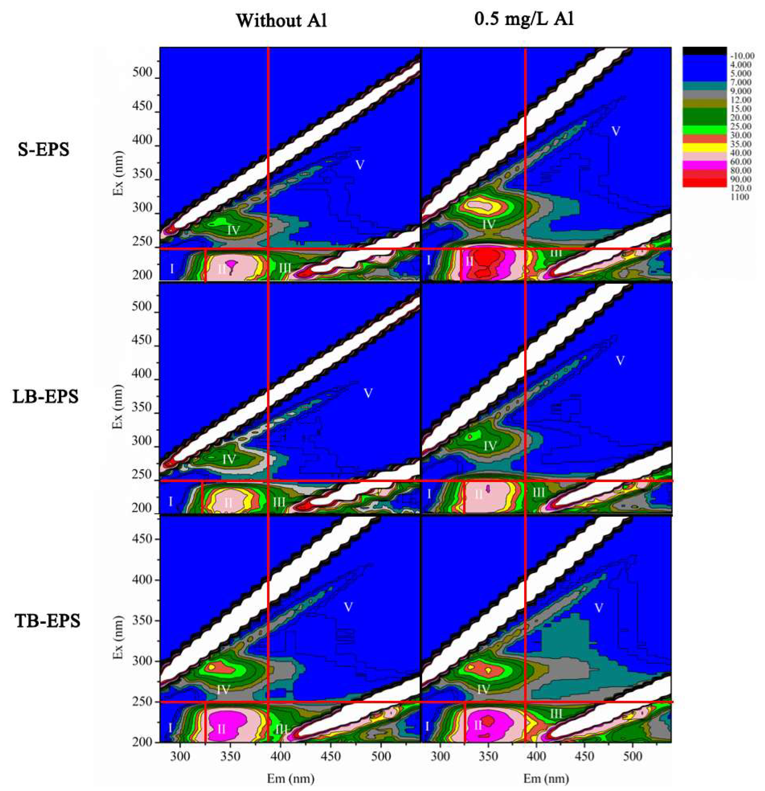
EPS was further evaluated utilizing the 3D-EEM fluorescence spectra (Figure 3). Two distinct peaks were clearly observed in the spectra of each EPS fraction; at 250–380/280–380 (Excition/Emition (Ex/Em)) (tryptophan-like or soluble microbial byproduct), and 220–250/320–380 (Ex/Em) (tyrosine/tryptophan protein). The EPS fractions content determined from the 3D-EEM fluorescence spectra increased with the aluminum ion concentration increase [24]. This is consistent with the EPS protein composition results.
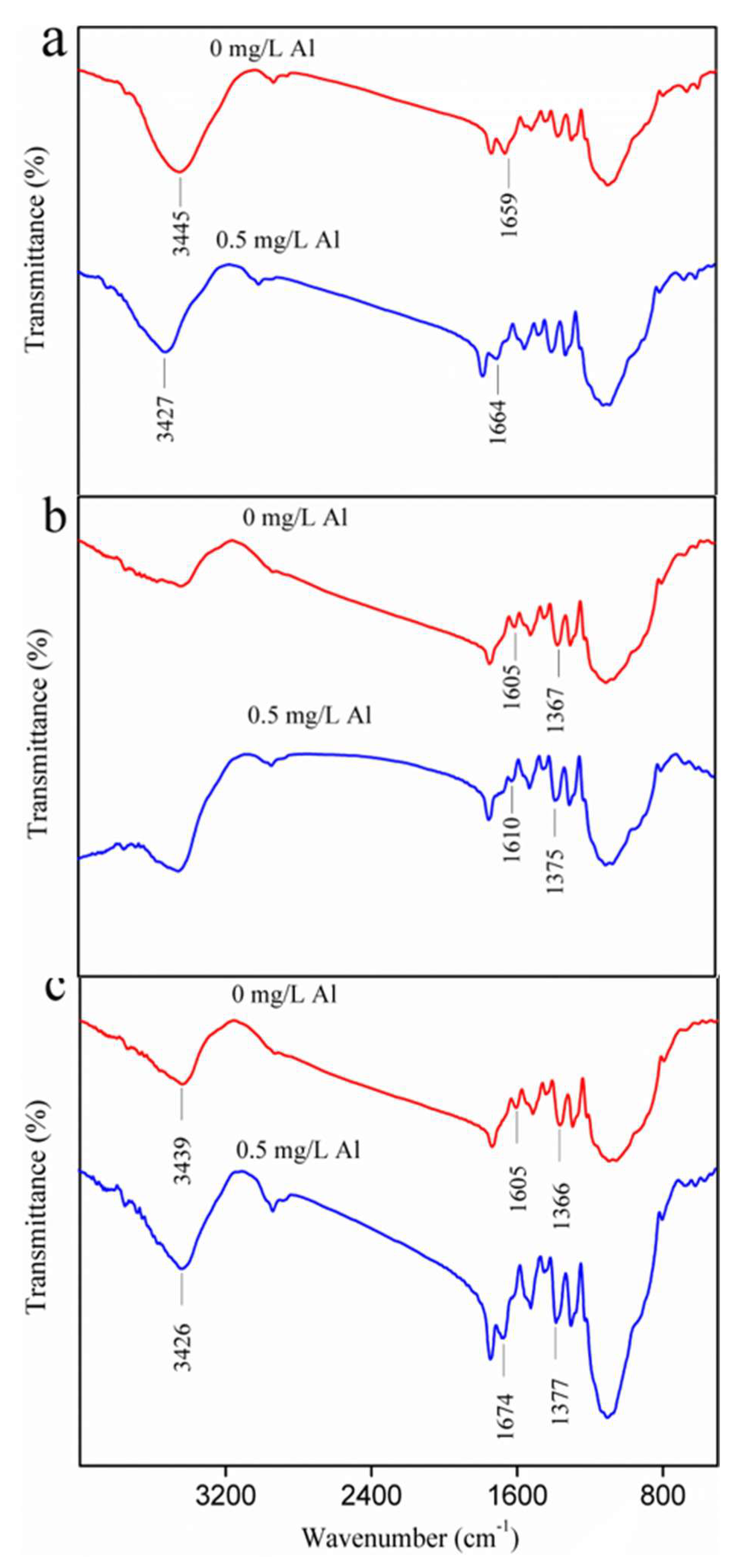
FTIR analysis over 4000–400 cm−1 was performed in order to identify changes in the EPS fraction functional groups following the exposure to aluminum ions (Figure 4). The broad absorption band near 3445 cm−1 is formed by the stretching vibration of both hydroxyl (of the carbohydrates) and amino groups (of the proteins) [26]. Moreover, the absorption band close to 1659 cm−1 is related to the stretching vibration of C = O in the presence of proteins [27]. The bands near 1610 cm−1 are attributed to the protein amide I region [28], while the band near 1375 cm−1 denotes the absorption of the C-H vibrations in the methyl. The broad adsorption peak around 3440 cm−1 was observed to have been impacted following aluminum exposure. This owes to the interaction of −OH and −NH2 with the aluminum ion. Variations between the FTIR spectra of S-EPS, LB-EPS and TB-EPS suggest that the different EPS functional groups were affected by aluminum ions, which may change the EPS physical characteristics, thus the zeta potential, flocculation efficiency, hydrophilicity and hydrophobicity were investigated.
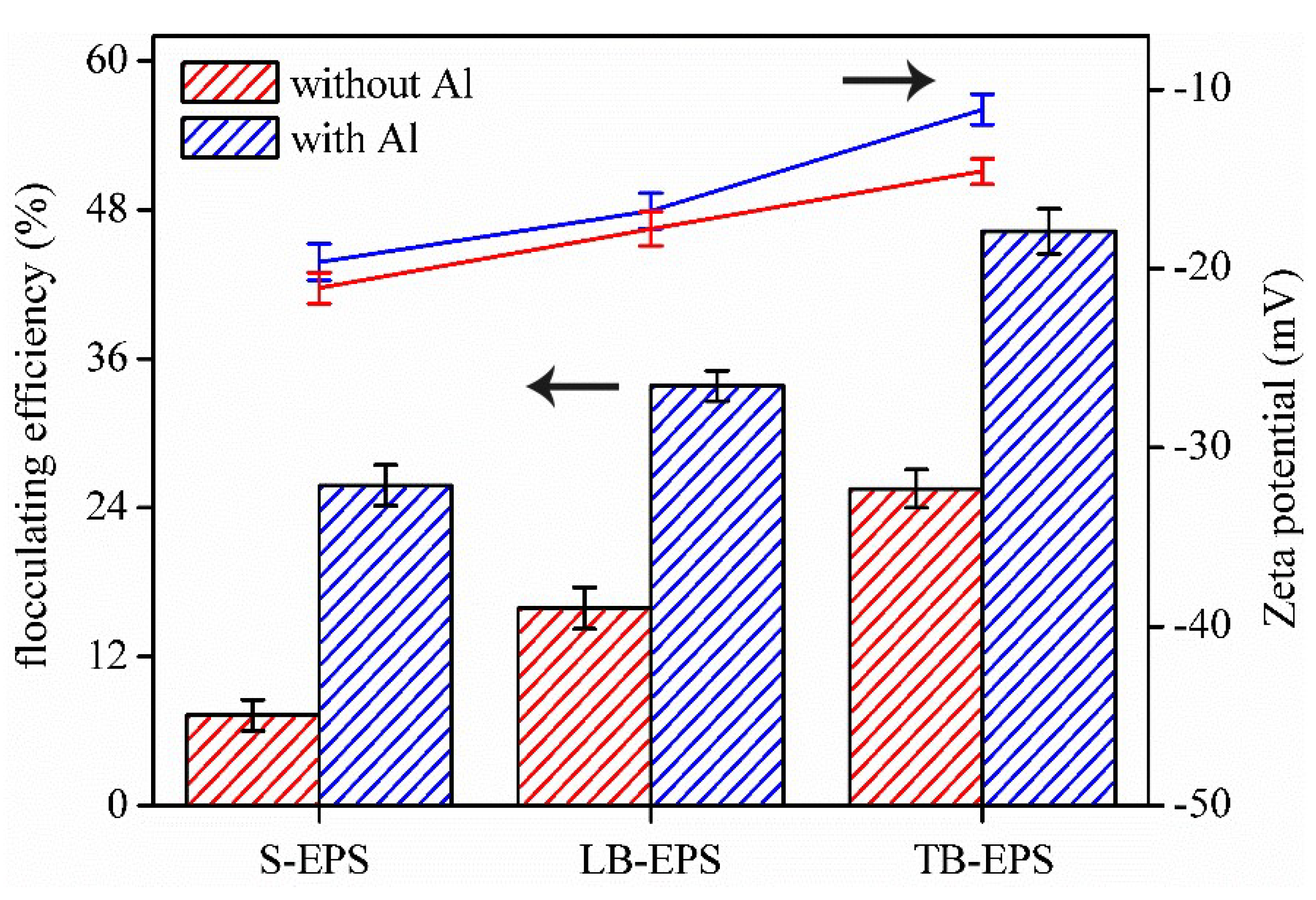
The observed zeta potential of each EPS fraction exhibited the following trend: S-EPS < LB-EPS < TB-EPS (Figure 5). In general, each EPS fraction exhibited a higher zeta potential than that of the control sample when exposed to 0.5 mg/L of aluminum ions. This is due to the neutralization and stabilization of the negative charge of the EPS functional groups with the addition of metal ions, thus increasing the zeta potential. Note that the flocculation efficiency of each EPS fraction agreed with the observed zeta potential trend (Figure 5). Flocculation experiments indicated that aluminum increased the flocculation efficiency and membrane fouling potential of each EPS fraction. This also means that the characteristics of EPS fractions were changed by the aluminum, which is consistent with the results of FTIR.
3.3. The Fouling Potential of the EPS Fractions
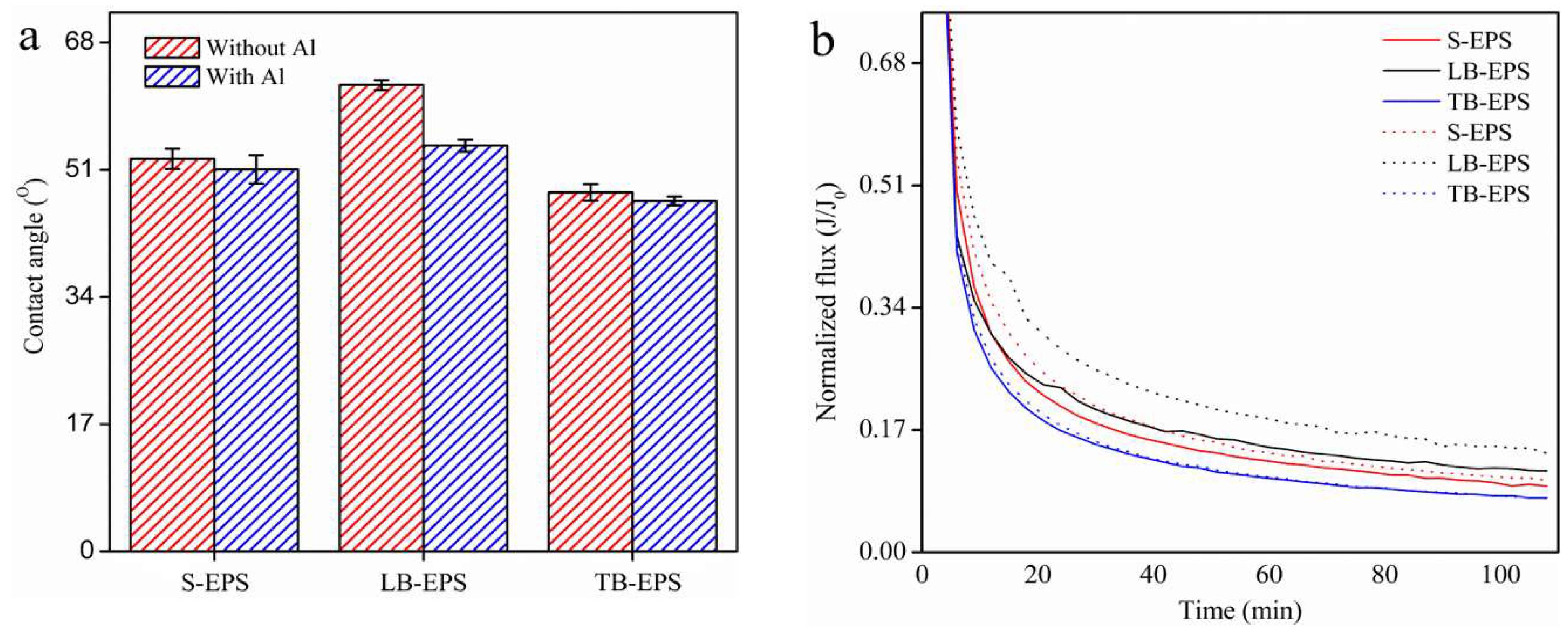
The hydrophilicity and hydrophobicity of the EPS fraction were determined using the water contact angle (Figure 6a). The contact angle of EPS cultured with aluminum was generally lower than that without aluminum. This indicates that the hydrophilicity of EPS decreased and hydrophobicity increased with the addition of aluminum ion, resulting in easier EPS depositions. Changes in hydrophilicity and hydrophobicity are related to the composition and content of the changed EPS via the aluminum ion binding with –OH and –COOH groups within the EPS [29]. The fouling potential results demonstrated that the fouling potential of S-EPS and LB-EPS was relatively smaller without aluminum (Figure 6b). The aluminum greatly increased the fouling potential of S-EPS and LB-EPS. Although TB-EPS has the highest fouling potential compared with that of S-EPS and LB-EPS, Al did not increase the TB-EPS fouling potential. Moreover, TB-EPS is the most stable part in biofilm, it does not detach from the carries easily (such as the distribution tubes), while S-EPS, LB-EPS could detach easily [30]. On account of this point, the increased fouling potential of S-EPS and LB-EPS by Al should receive more attention.
4. Conclusions
Different biofilm fractions have different amounts of residual Al. This study first revealed that residual Al significantly increased the membrane fouling potentials of both S-EPS and LB-EPS rather than that of TB-EPS. These increasing membrane fouling potentials could be explained by the increasing zeta potential and flocculation ability of the EPS in biofilm because of residual Al. The residual Al in biofilm induced more biofilm formation and more EPS formation, which could also induce serious membrane fouling. Al coagulants should be concerned in wastewater reclamation. In future studies, an effective coagulant should be found in order to decrease the membrane fouling potentials of biofilm in wastewater reclaiming.
Author Contributions
Conceptualization, X.C.; Data curation, Z.G.; Formal analysis, S.S. and Z.Z.; Funding acquisition, X.C.; Methodology, Z.G.; Supervision, X.C. and M.H.; Writing—original draft, S.S.; Writing—review & editing, X.C. All authors have read and agreed to the published version of the manuscript.
Funding
The authors wish to thank the National Natural Science Foundation of China (51808103, 51578117 and 51508079) and the Fundamental Research Funds for the Central Universities (2412018QD021) for their financial support.
Conflicts of Interest
There is no conflict of interest.
References
- Bellona, C.; Drewes, J.E. Viability of a low-pressure nanofilter in treating recycled water for water reuse applications: A pilot-scale study. Water Res. 2007, 41, 3948–3958. [Google Scholar] [CrossRef] [PubMed]
- Schneider, R.P.; Ferreira, L.M.; Binder, P.; Ramos, J.R. Analysis of foulant layer in all elements of an RO train. J. Membr. Sci. 2005, 261, 152–162. [Google Scholar] [CrossRef]
- Huang, S.; Voutchkov, N.; Jiang, S. Balancing carbon, nitrogen and phosphorus concentration in seawater as a strategy to prevent accelerated membrane biofouling. Water Res. 2019, 165, 114978. [Google Scholar] [CrossRef] [PubMed]
- Matin, A.; Khan, Z.; Zaidi, S.; Boyce, M. Biofouling in reverse osmosis membranes for seawater desalination: Phenomena and prevention. Desalination 2011, 281, 1–16. [Google Scholar] [CrossRef]
- Julien, C.; Laurent, E.; Legube, B.; Thomassin, J.-H.; Mondamert, L.; Labanowski, J. Investigation on the iron-uptake by natural biofilms. Water Res. 2014, 50, 212–220. [Google Scholar] [CrossRef] [Green Version]
- You, G.; Hou, J.; Xu, Y.; Wang, C.; Wang, P.; Miao, L.; Ao, Y.; Li, Y.; Lv, B. Effects of CeO2 nanoparticles on production and physicochemical characteristics of extracellular polymeric substances in biofilms in sequencing batch biofilm reactor. Bioresour. Technol. 2015, 194, 91–98. [Google Scholar] [CrossRef]
- Zhang, W.; Cao, B.; Wang, D.; Ma, T.; Xia, H.; Yu, D. Influence of wastewater sludge treatment using combined peroxyacetic acid oxidation and inorganic coagulants re-flocculation on characteristics of extracellular polymeric substances (EPS). Water Res. 2016, 88, 728–739. [Google Scholar] [CrossRef]
- Desmond, P.; Best, J.P.; Morgenroth, E.; Derlon, N. Linking composition of extracellular polymeric substances (EPS) to the physical structure and hydraulic resistance of membrane biofilms. Water Res. 2018, 132, 211–221. [Google Scholar] [CrossRef]
- Zhao, H.; Liu, H.; Hu, C.; Qu, J. Effect of aluminum speciation and structure characterization on preferential removal of disinfection byproduct precursors by aluminum hydroxide coagulation. Environ. Sci. Technol. 2009, 43, 5067–5072. [Google Scholar] [CrossRef]
- Cui, X.; Zhou, D.; Fan, W.; Huo, M.; Crittenden, J.C.; Yu, Z.; Ju, P.; Wang, Y. The effectiveness of coagulation for water reclamation from a wastewater treatment plant that has a long hydraulic and sludge retention times: A case study. Chemosphere 2016, 157, 224–231. [Google Scholar] [CrossRef]
- Cui, X.; Huo, M.; Chen, C.; Yu, Z.; Zhou, C.; Li, A.; Qiao, B.; Zhou, D.; Crittenden, J.C. Low concentrations of Al (III) accelerate the formation of biofilm: Multiple effects of hormesis and flocculation. Sci. Total Environ. 2018, 634, 516–524. [Google Scholar] [CrossRef] [PubMed]
- Li, B.; Huang, W.; Zhang, C.; Feng, S.; Zhang, Z.; Lei, Z.; Sugiura, N. Effect of TiO2 nanoparticles on aerobic granulation of algal–bacterial symbiosis system and nutrients removal from synthetic wastewater. Bioresour. Technol. 2015, 187, 214–220. [Google Scholar] [CrossRef] [PubMed]
- Bajaj, M.; Gallert, C.; Winter, J. Biodegradation of high phenol containing synthetic wastewater by an aerobic fixed bed reactor. Bioresour. Technol. 2008, 99, 8376–8381. [Google Scholar] [CrossRef] [PubMed]
- You, G.; Wang, P.; Hou, J.; Wang, C.; Xu, Y.; Miao, L.; Lv, B.; Yang, Y.; Liu, Z.; Zhang, F. Insights into the short-term effects of CeO2 nanoparticles on sludge dewatering and related mechanism. Water Res. 2017, 118, 93–103. [Google Scholar] [CrossRef]
- Raunkjær, K.; Hvitved-Jacobsen, T.; Nielsen, P.H. Measurement of pools of protein, carbohydrate and lipid in domestic wastewater. Water Res. 1994, 28, 251–262. [Google Scholar] [CrossRef]
- Zhang, P.; Fang, F.; Chen, Y.-P.; Shen, Y.; Zhang, W.; Yang, J.-X.; Li, C.; Guo, J.-S.; Liu, S.-Y.; Huang, Y. Composition of EPS fractions from suspended sludge and biofilm and their roles in microbial cell aggregation. Chemosphere 2014, 117, 59–65. [Google Scholar] [CrossRef]
- Wilén, B.-M.; Lumley, D.; Mattsson, A.; Mino, T. Relationship between floc composition and flocculation and settling properties studied at a full scale activated sludge plant. Water Res. 2008, 42, 4404–4418. [Google Scholar] [CrossRef]
- Yu, T.; Li, G.; Lin, W.; Hu, H.-Y.; Lu, Y. Coagulation increased the growth potential of various species bacteria of the effluent of a MBR for the treatment of domestic wastewater. Environ. Sci. Pollut. Res. 2017, 24, 5126–5133. [Google Scholar] [CrossRef]
- Hou, J.; You, G.; Xu, Y.; Wang, C.; Wang, P.; Miao, L.; Li, Y.; Ao, Y.; Lv, B.; Yang, Y. Long-term effects of CuO nanoparticles on the surface physicochemical properties of biofilms in a sequencing batch biofilm reactor. Appl. Microbiol. Biotechnol. 2016, 100, 9629–9639. [Google Scholar] [CrossRef]
- Flemming, H.-C.; Wingender, J. The biofilm matrix. Nat. Rev. Microbiol. 2010, 8, 623–633. [Google Scholar] [CrossRef]
- Wang, B.-B.; Chang, Q.; Peng, D.-C.; Hou, Y.-P.; Li, H.-J.; Pei, L.-Y. A new classification paradigm of extracellular polymeric substances (EPS) in activated sludge: Separation and characterization of exopolymers between floc level and microcolony level. Water Res. 2014, 64, 53–60. [Google Scholar] [CrossRef]
- Laspidou, C.S.; Rittmann, B.E. A unified theory for extracellular polymeric substances, soluble microbial products, and active and inert biomass. Water Res. 2002, 36, 2711–2720. [Google Scholar] [CrossRef]
- Cui, X.; Chen, C.; Liu, Y.; Zhou, D.; Liu, M. Exogenous refractory protein enhances biofilm formation by altering the quorum sensing system: A potential hazard of soluble microbial proteins from WWTP effluent. Sci. Total Environ. 2019, 667, 384–389. [Google Scholar] [CrossRef]
- Zhang, Z.-J.; Chen, S.-H.; Wang, S.-M.; Luo, H.-Y. Characterization of extracellular polymeric substances from biofilm in the process of starting-up a partial nitrification process under salt stress. Appl. Microbiol. Biotechnol. 2011, 89, 1563–1571. [Google Scholar] [CrossRef]
- Chen, W.; Westerhoff, P.; Leenheer, J.A.; Booksh, K. Fluorescence excitation—Emission matrix regional integration to quantify spectra for dissolved organic matter. Environ. Sci. Technol. 2003, 37, 5701–5710. [Google Scholar] [CrossRef]
- Zhu, L.; Dai, X.; Zhou, J.-h.; Xu, X.-y. The stability of aerobic granular sludge under 4-chloroaniline shock in a sequential air-lift bioreactor (SABR). Bioresour. Technol. 2013, 140, 126–130. [Google Scholar] [CrossRef]
- Liang, Z.; Li, W.; Yang, S.; Du, P. Extraction and structural characteristics of extracellular polymeric substances (EPS), pellets in autotrophic nitrifying biofilm and activated sludge. Chemosphere 2010, 81, 626–632. [Google Scholar] [CrossRef]
- Yin, C.; Meng, F.; Chen, G.-H. Spectroscopic characterization of extracellular polymeric substances from a mixed culture dominated by ammonia-oxidizing bacteria. Water Res. 2015, 68, 740–749. [Google Scholar] [CrossRef] [PubMed]
- Sheng, G.-P.; Yu, H.-Q. Relationship between the extracellular polymeric substances and surface characteristics of Rhodopseudomonas acidophila. Appl. Microbiol. Biotechnol. 2006, 72, 126–131. [Google Scholar] [CrossRef] [PubMed]
- Wang, J.; Liu, Q.; Hu, H.; Wu, B.; Zhang, X.-x.; Ren, H. Insight into mature biofilm quorum sensing in full-scale wastewater treatment plants. Chemosphere 2019, 234, 310–317. [Google Scholar] [CrossRef] [PubMed]
Figure 1.
The influence of Aluminum on (a) biofilm dry weight and (b) extracellular polymeric substances (EPS) fraction content.
Figure 1.
The influence of Aluminum on (a) biofilm dry weight and (b) extracellular polymeric substances (EPS) fraction content.

Figure 2.
The spatial distribution of Al in biofilm (a) and the influence of Al on EPS fraction composition (soluble EPS (S-EPS), loosely bound EPS (LB-EPS) and tightly bound (TB-EPS)) (b).
Figure 2.
The spatial distribution of Al in biofilm (a) and the influence of Al on EPS fraction composition (soluble EPS (S-EPS), loosely bound EPS (LB-EPS) and tightly bound (TB-EPS)) (b).

Figure 3.
Excitation-emission matrix EEM fluorescence spectra of EPS extracted from the biofilm by cultured 0 mg/L Al (left: S-EPS, LB-EPS, TB-EPS) and cultured by 0.5 mg/L Al (right: S-EPS, LB-EPS, TB-EPS). Five regions were classified according to the methods of Chen (2003) [25] as follows. Region I denotes the tyrosine/tryptophan amino acid region with Ex/Em = 220–250/280–320, region II denotes the tyrosine/tryptophan protein region with Ex/Em = 220–250/320–380, region III denotes the fulvic acid region with Ex/Em = 220–250/380–460, region IV denotes the tryptophan-like or soluble microbial byproduct-like material with Ex/Em = 250–380/280–380, and region V denotes humic acid-like organics with Ex/Em = 250–380/380–460.

Figure 4.
Fourier-transform infrared spectrophotometer (FTIR) spectra of the EPS fractions ((a) S-EPS, (b) LB-EPS and (c) TB-EPS) extracted from biofilms cultivated with and without Al.
Figure 4.
Fourier-transform infrared spectrophotometer (FTIR) spectra of the EPS fractions ((a) S-EPS, (b) LB-EPS and (c) TB-EPS) extracted from biofilms cultivated with and without Al.

Figure 5.
Zeta potential and flocculation efficiency of each EPS fraction.

Figure 6.
Contact angle of EPS fractions with and without aluminum (a) and normalized flux reduction of EPS fractions after being dissolved to the EPS concentration of 0.1 mg/L. Solid line represents the groups with Al, while dashed line represents the groups without Al (b).
Figure 6.
Contact angle of EPS fractions with and without aluminum (a) and normalized flux reduction of EPS fractions after being dissolved to the EPS concentration of 0.1 mg/L. Solid line represents the groups with Al, while dashed line represents the groups without Al (b).

© 2020 by the authors. Licensee MDPI, Basel, Switzerland. This article is an open access article distributed under the terms and conditions of the Creative Commons Attribution (CC BY) license (http://creativecommons.org/licenses/by/4.0/).
Share and Cite
MDPI and ACS Style
Sun, S.; Zhao, Z.; Cui, X.; Huo, M.; Geng, Z. The Influence of Residual Coagulant Al on the Biofilm EPS and Membrane Fouling Potential in Wastewater Reclamation. Water 2020, 12, 1056. https://doi.org/10.3390/w12041056
AMA Style
Sun S, Zhao Z, Cui X, Huo M, Geng Z. The Influence of Residual Coagulant Al on the Biofilm EPS and Membrane Fouling Potential in Wastewater Reclamation. Water. 2020; 12(4):1056. https://doi.org/10.3390/w12041056
Chicago/Turabian StyleSun, Shu, Zhenhao Zhao, Xiaochun Cui, Mingxin Huo, and Zhi Geng. 2020. "The Influence of Residual Coagulant Al on the Biofilm EPS and Membrane Fouling Potential in Wastewater Reclamation" Water 12, no. 4: 1056. https://doi.org/10.3390/w12041056
Note that from the first issue of 2016, this journal uses article numbers instead of page numbers. See further details here.