The Use of Boron-Doped Diamond Electrode for the Determination of Selected Biocides in Water Samples


Institute of Chemistry, University of Białystok, Ciołkowskiego 1 K, 15-245 Białystok, Poland
*
Author to whom correspondence should be addressed.
Water 2019, 11(8), 1595; https://doi.org/10.3390/w11081595
Submission received: 24 June 2019
/
Revised: 16 July 2019
/
Accepted: 30 July 2019
/
Published: 31 July 2019
(This article belongs to the Special Issue Removal of Organic Pollution in Water Environment)
Abstract
:In recent years, the remains of chemical substances in water environments, referred to as emerging organic contaminations, have been more and more often studied by analysts. This work shows the possibility of using a boron-doped diamond electrode to determine low concentration levels of remains of pharmaceuticals in environmental samples. The study focused on selected biocides from the group of azole fungicides (itraconazole and posaconazole) and was performed using quick and sensitive electrochemical methods. The cyclic voltammetry method was used in order to determine the properties of these compounds, whereas analytical characterization was performed using square wave voltammetry. The work involved the specification of the optimum electrooxidation conditions of the selected fungicides, their comparative characterization, and the development of a new, sensitive methods of itraconazole and posaconazole assay. The proposed procedures allowed us to determine itraconazole in the range from 7.9 × 10⁻⁸ to 1.2 × 10⁻⁶ moL·L−1 and posaconazole in the range from 5.7 × 10⁻⁸ to 8.44 × 10⁻⁷ moL·L−1. The relative standard deviation of the measurements did not exceed 5.85%. The developed procedures were successfully used to determine itraconazole and posaconazole concentration in water samples and the assay recovery was between 93.5% and 102.8%.
1. Introduction
The issue of remains of pharmaceutical substances in water environments is becoming a global problem. The development of the pharmaceutical industry, the advancement of medicine, the increasing number of civilization diseases, the appearance of antibiotic-resistant bacteria and the growing consumption of drugs as a part of disease prevention lead to a dramatic growth in the amount of pharmaceutical contamination in water and wastewater. Particular attention is given to the remains of chemicals referred to as “emerging organic contaminants”, such as active ingredients of pharmaceuticals, cosmetics, preservatives, and surfactants. In surface water, wastewater, and even drinking water, the remains of medicines and their active metabolites are more and more often found in amounts expressed in ng/L or µg/L. Water contaminated by them is a serious threat for the life and health of humans and animals. Classical methods of water purification are not effective in eliminating the broad spectrum of newly emerging pharmaceuticals, which get to drinking water, groundwater or bottom sediments. Therefore, studies related to the environmental impact assessment of pharmaceuticals are needed. It is necessary to develop analytical methods for different environmental matrices. The development of analytical methods for validation of pharmaceuticals in environmental matrices is becoming more and more important and necessary [1,2,3,4,5,6].
One group of compounds that have been given more and more attention recently is biocides [7]. Water contamination with these compounds results from their common use in daily life. They are used as active substances in pharmaceutical preparations or body care products (e.g., creams, ointments or shampoos) [8]. The presence of biocides has already been observed in sewage treatment plants and various environmental media [9,10,11,12,13]. For example, miconazole, ketoconazole and fluconazole have been detected in wastewater in concentrations up to 36, 90 and 140 ng/mL, respectively [14]. Itraconazole, fenticonazole and tioconazole have also been detected in sludge from a sewage treatment plant in real samples from the Northwest of Spain in concentrations of 204, 110 and 74 ng/g, respectively [15]. Pharmaceuticals in environmental samples are usually detected using gas or liquid chromatography methods, sometimes combined with tandem mass spectrometry [16]. Spectroscopic methods such as near infrared spectroscopy (NIR) or nuclear magnetic resonance (NMR) are also used [17].
Electrochemical methods and boron-doped diamond electrodes (BDD) are more and more often used to assay pharmaceuticals in environmental samples. The BDD is a new electrode material, effective also in the degradation and removal of contamination from water samples [18,19]. The BDD electrode allows us to measure the analyzed samples quickly, and the low and stable current ensures the high sensitivity of the measurements. Thanks to its unique physical and chemical properties [20,21,22,23,24], the boron-doped diamond electrode is an alternative to traditional carbon electrodes. The BDD electrode ensures excellent chemical resistance and stability in water environments. It has a very wide potential window, so it can be used in various electrochemical reactions in water environment. In addition, it has poor adsorption properties and high ability of oxidizing organic and inorganic compounds [25,26,27,28,29,30]. A conductive diamond has been used to assay biologically active compounds in various water matrices. This electrode has been used, in other words, to assay chemotherapeutics (e.g., ciprofloxacin) in natural waters and wastewater [31]. The BDD electrode has been used to assay various antibiotics (tetracycline, erythromycin, sulfamethoxazole), antidepressants (e.g., fluoxetine, viloxazine) and analgesics (e.g., naproxen) in environmental water samples [32,33,34,35,36].
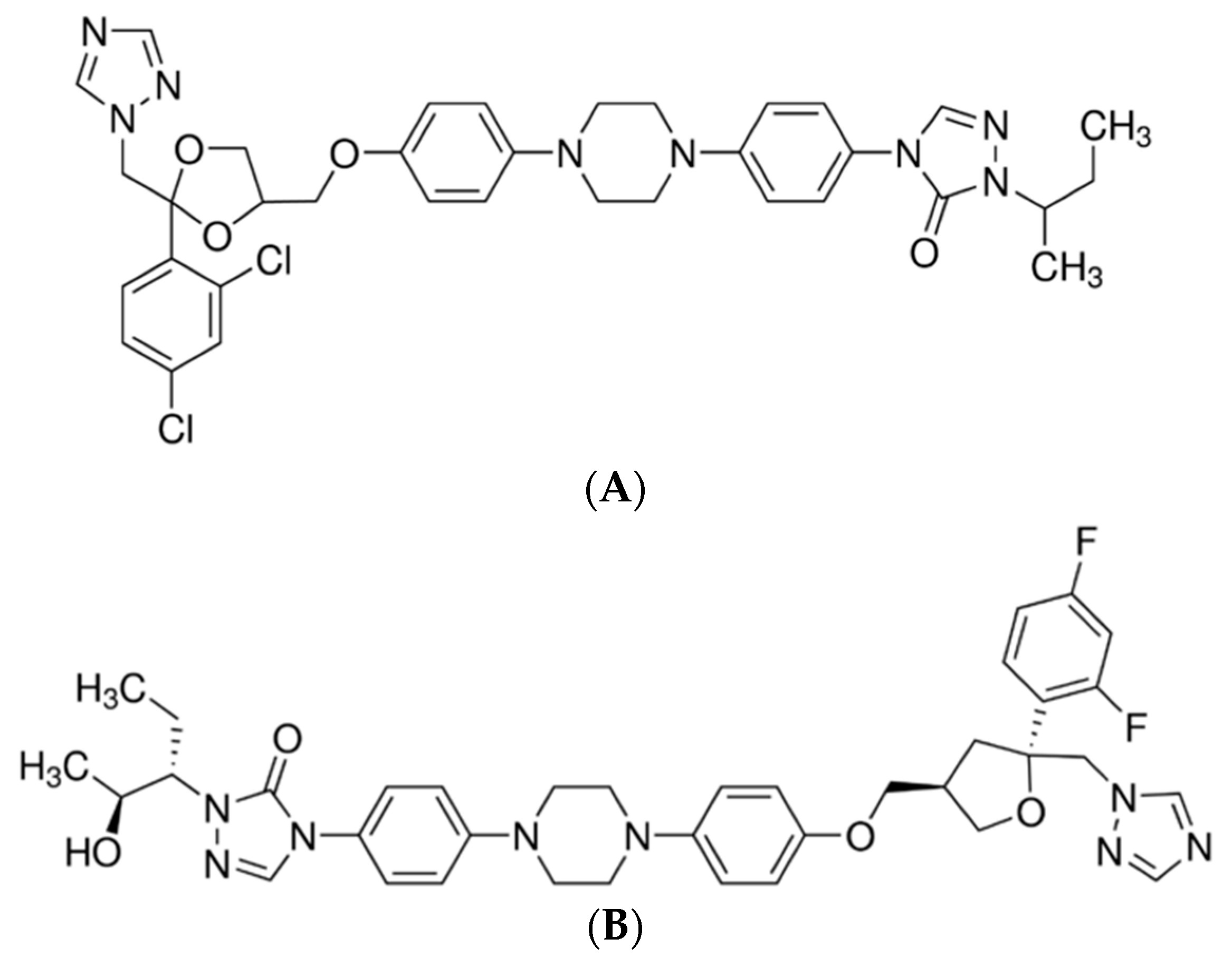
In this work the use of the BDD electrode to assay selected biocides in water samples contaminated with pharmaceutical substances is proposed (Figure 1).
2. Experimental
Apparatus and Chemicals
All the experiments were carried out using the Autolab PGSTAT 128N potentiostat from Metrohm Autolab B.V. with NOVA ver. 1.10 software, allowing for computer data collection and analysis of the results. The set used in voltammetric measurements included three electrodes. The working electrode was a D-035-SA boron-doped diamond electrode (BDD) from Windsor Scientific LTD, Slough, UK (diamond doped with boron, around 0.1%, A = 0.07 cm2). The auxiliary electrode was a platinum plate with the surface area A = 0.9 cm2. The reference electrode was EK-602 saturated calomel electrode (SCE) from Eurosensor.
Before each measurement series, the surface of BDD electrode was activated through cathodic reduction of hydrogen within the potential range of −2.9 V to 0.3 V. In the case of adsorption of the studied substance on the surface of the working electrode, it was mechanically purified using an MF-2060 polishing cloth from Bioanalytical System (USA) covered with aluminum oxide with grain sizes of 0.05 µm or 0.01 µm. All the experiments were carried out at room temperature and in the presence of oxygen. The studied solutions were not deoxidized before the voltammetric measurement.
In order to prepare the solutions, the analytes were weighted on a PB-153 scale (Mettler Toledo, Greifensee, Switzerland). The solutions’ pH was measured with a pH-meter inoLab Level 1 from WTW, Germany. All the substances used in the experiments—itraconazole (ITR), posaconazole (POZ)—were purchased from Sigma-Aldrich, Hamburg, Germany. The solution of posaconazole with the concentration of 5.7 × 10⁻⁴ mol/L and the solution of itraconazole with the concentration of 1 × 10⁻³ mol/L were prepared by dissolving the appropriate weighted amounts of those substances in methanol. All the solutions were kept in a refrigerator.
The supporting electrolytes were 0.1 mol/L solution of potassium chloride, Britton-Robinson (B-R) buffer solutions with pH: 1.81; 2.29; 2.87; 4.35; 5.33; 7.0; 8.36; 9.15; 10.38; 11.20. B-R solutions with these pH values were prepared by mixing 0.4 mol/L H3PO4, 0.04 mol/L H3BO3 and 0.04 mol/L CH3COOH with the appropriate amount of 0.2 mol/L NaOH.
The interferents were substances that may potentially occur in water, such as salts of sodium, potassium, iron(II), magnesium, copper, lead, calcium, cadmium and zinc, ions: Cl−, SO42−, NO3−, organic substances: Triton X-100, sodium dodecyl sulfate (SDS), tetrabutylamonium bromide, ketoconazole, clotrimazole, voriconazole, triclosan and methylparaben. The solutions of these salts with the concentrations of 2.8 × 10⁻⁴ mol/L were prepared by dissolving the appropriate weighted amounts of relevant substances in water. All the solutions were prepared using deionized water Milli–Q (Millipore Corp., Burlington, MA, USA).
3. Results and Discussion
3.1. Electrochemical Behavior of Selected Biocides
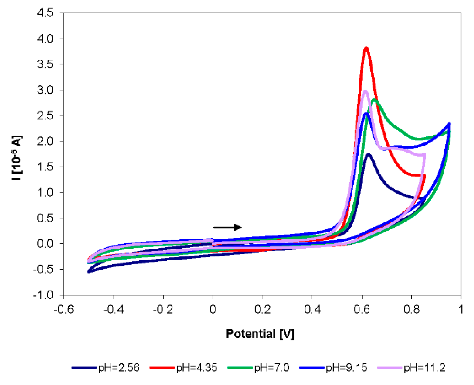
Azoles are the most popular and the most frequently used class of fungicides. First-generation azoles are imidazoles (e.g., ketoconazole), while second- and third-generation azoles are triazoles (e.g., posaconazole and itraconazole studied in the work). The selected azoles were studied using cyclic voltammetry (CV). In order to determine the optimum conditions of oxidation of selected analytes, the influence of the kind and pH of the used supporting electrolyte was studied. The experiments were carried out in Britton-Robinson buffer in the pH range of 1.8 to 11.2. Figure 2 presents the cyclic curves plotted in various buffer solutions for posaconazole.
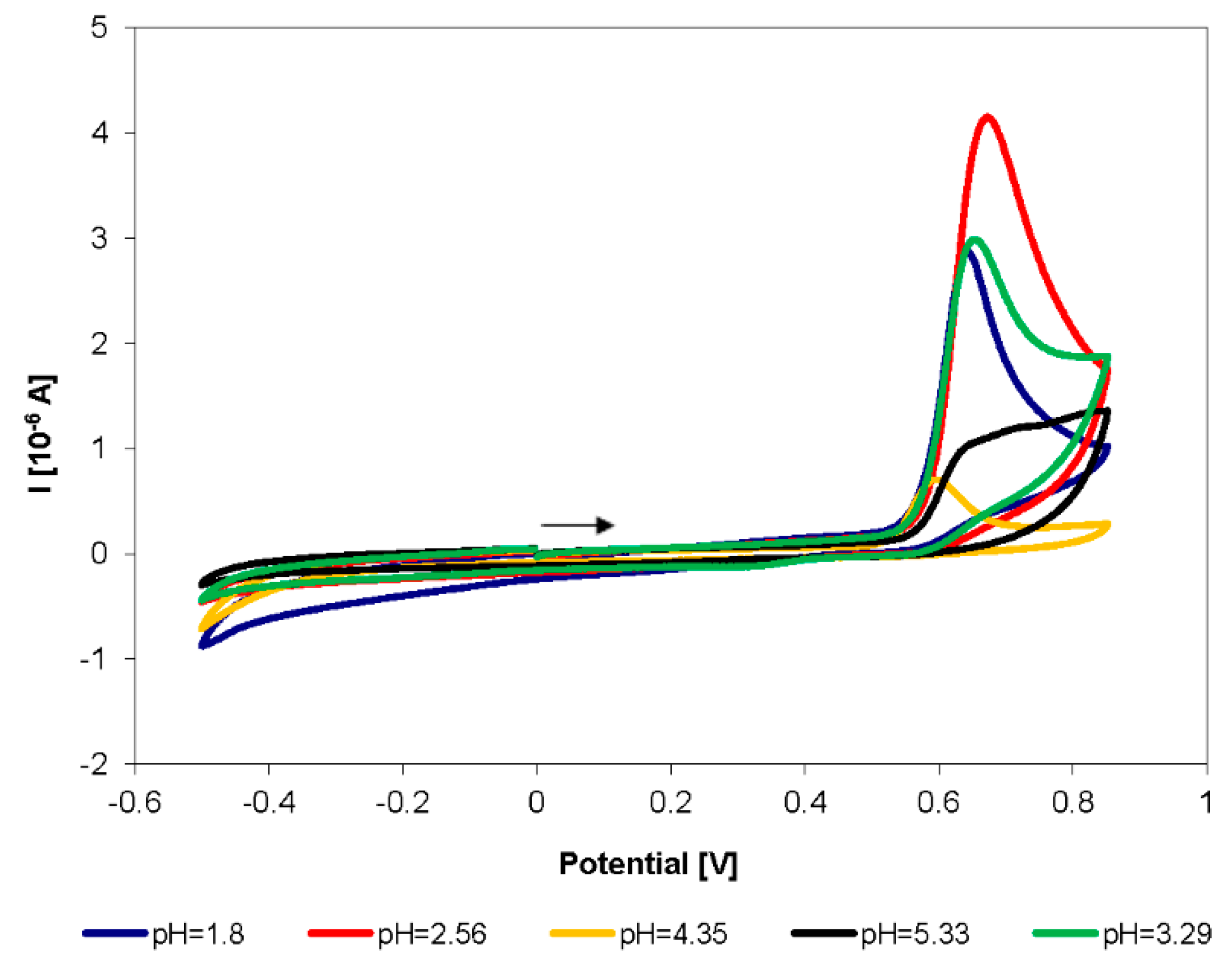
Analogically, oxidation curves of itraconazole as a compound structurally similar to posaconazole were plotted (Figure 3).
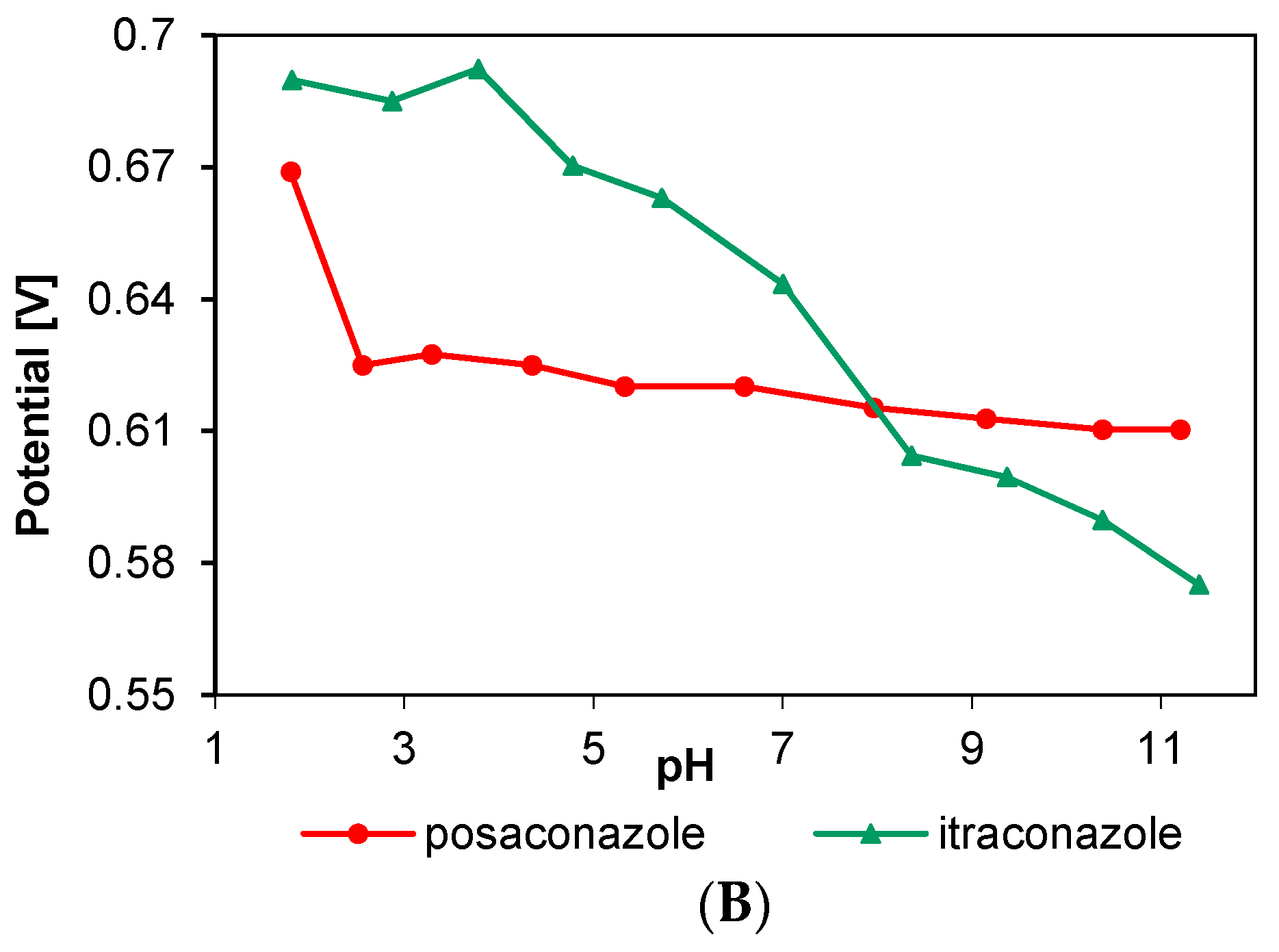
The voltammograms of all the studied compounds showed irreversible oxidation curves in the potential range of 0.58 V do 0.70 V. Figure 2 and Figure 3 show example curves from all the studied period, and the complete characteristics of relationships between current and oxidation peak potential depending on pH are presented in Figure 4.
The highest values of itraconazole oxidation peak were obtained in the B-R buffer with pH = 2.87, and of posaconazole in the solution with pH = 4.35. As the pH of the electrolyte increased, the potentials of recorded peaks of itraconazole and posaconazole moved towards lower values, and the current values decreased. The obtained results confirm literature data and earlier studies for ketoconazole [26,37]. The analysis of the above-mentioned azoles depending of the pH of the solution showed that the compounds’ structure determines their electrochemical behavior. The comparable electrochemical behavior of these azole fungicides may prove the similarity of electrochemical reactions.
The most probable mechanism of oxidation of these compounds is the loss of an electron in the piperazine ring. Similar conclusions were also formulated for other substances containing a piperazine ring [38,39]. That oxidation produced lower currents with decreasing pH values. Considering the values: pKa, 3.6 for posaconazole and 3.7 for itraconazole [40,41], as well as the pH-dependence, the deprotonated piperazine group can be regarded as an electroactive form. This is also proved by the shift in the peak potential with the growth of pH. It shows that the deprotonated piperazine group must be produced by the dissociation of the proton [42].
3.2. Effect of Scan Rate
In order to determine the character of the recorded currents, the impact of the scan rate on the values of recorded currents was investigated. The experiments were carried out at the scan rates: 5, 10, 25, 50, 75, 100, 200, 350 and 500 mV/s in the range of potentials from −0.2 V to 0.95 V. The measurements were performed in Britton-Robinson buffer solutions with properly selected pH values at which the highest current values were recorded for different analytes.
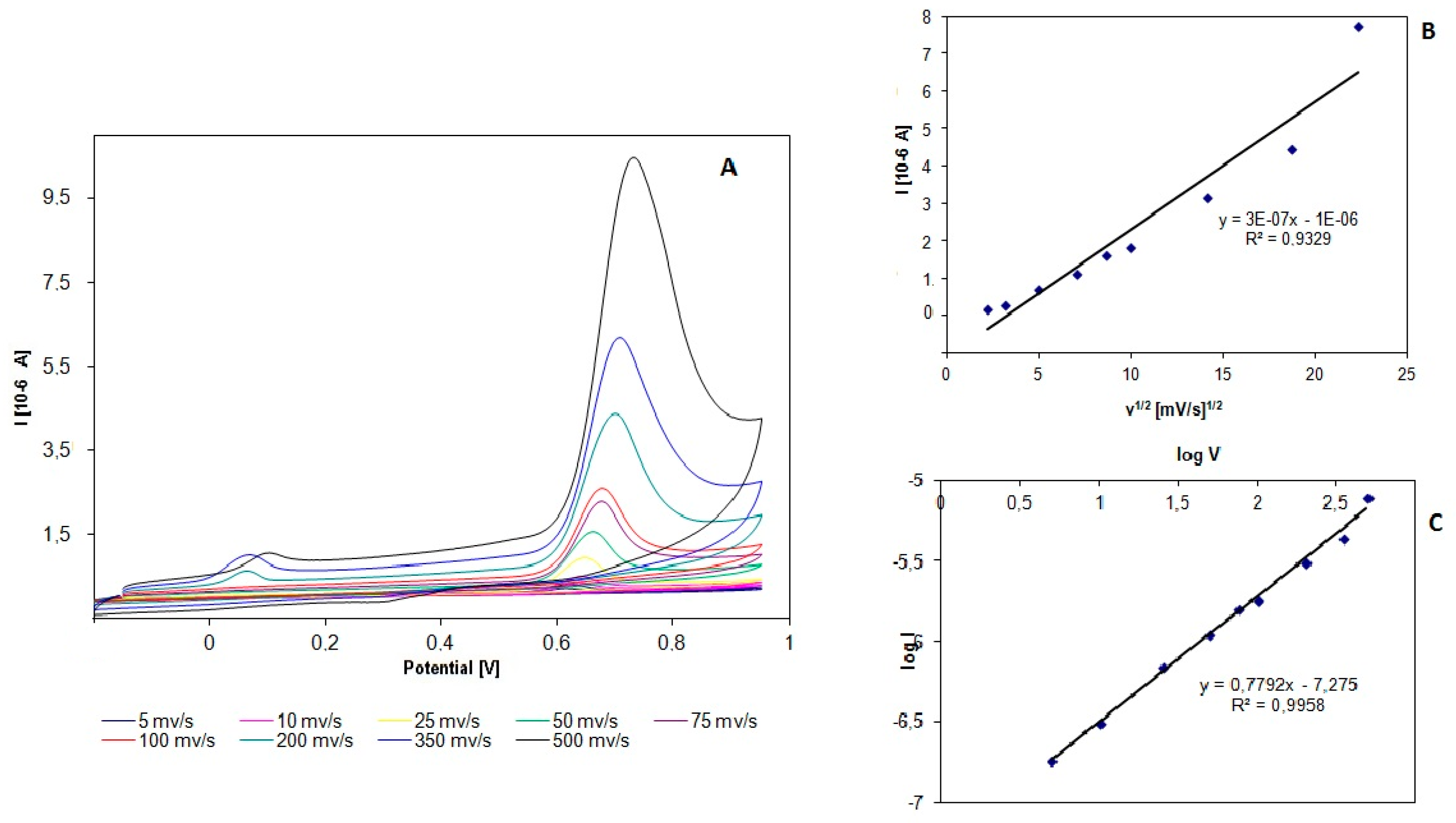
The cyclic curves of itraconazole recorded at different scan rates are shown in Figure 5.
The increase of the values of recorded currents along the increase of scan rate was observed for all the studied compounds. In the case of measurements for posaconazole, the analysis of peak current values of the studied compound depending on the root of the scan rate displayed a diffusion character. In the case of itraconazole, a clear decrease of the recorded current values was observed in successive cycles. This may be the evidence for itraconazole depositing on the surface of the electrode. The analysis of the logarithm of peak current of scan rate confirmed the diffusion character of currents in the case of posaconazole. The slope in the equations of curves was close to the theoretical value: 0.58 [43]. In the case of itraconazole, the slope of the curve was 0.77, which proves the mixed, diffusion and adsorption character of the measured currents. Figure 5C shows the relationship between the change in the logarithm of current and the logarithm of scan rate for the studied azole (itraconazole).
The equations describing these relationships can have the following form:
- for itraconazole (ITR)I (µA) = 3·10−7 v1/2 (mVs−1)1/2 − 1·10−6
- for posaconazole (POZ)I (µA) = 9·10−8 v1/2 (mVs−1)1/2 − 1·10−7
Based on the data from the obtained curves, the following equations were produced:
- ITRlog Ipa (µA) = 0.7792 log v(mV/s) − 7275, R2 = 0.9958
- POZlog Ipa (µA) = 0.58 log v(mV/s) − 7295, R2 = 0.9996
3.3. Optimization of the Square Wave Parameters
The square wave voltammetry (SWV) technique was chosen to develop the procedure of assaying a compound from the group of azoles (itraconazole). It is one of the most sensitive electrochemical techniques, ensuring the high sensitivity of assays. The experimental parameters characteristic of SWV were determined as part of the work. The optimization of the procedure involved the amplitude, frequency and step potential. The amplitude was studied in the range of potentials from 5 to 100 mV. The measurements were performed at constant values of frequency and step potential. The increase in the amplitude value caused the increase in recorded currents, transition of peak potentials toward negative values, but also increased width of the peaks. Therefore, the optimum value of amplitude selected for further experiments is 25 mV. The impact of frequency on the height of recorded peaks was checked for the values: 8, 15, 25, 50, 75, 100, 125 and 150 Hz. The increase in the measured current was observed in the whole studied range. A linear relationship of current depending on the root of frequency was observed for the range of 8 Hz to 100 Hz. Hence, the value of 100 Hz was adopted as the best for further experiments. The study of the impact of step potential on the value of oxidation current of itraconazole showed that, as this parameter increased in the range of 2 mV to 10 mV, the recorded currents decreased. The value of 2 mV/s was chosen for further study. All the measurements were carried out in the B-R buffer solution with pH = 2.87 in the potential range of −0.2 V to 0.95 V.
The same procedure was carried out for posaconazole. The experiments led to the determination of the optimum amplitude value. Just like in the study of itraconazole, the best values were obtained for 25 mV. Testing frequencies between 8 and 150 Hz led to the choice of the optimum value of this parameter. The value of 100 Hz was chosen for the study, because as the frequency increased, the width of the recorded peaks also grew. The tests of step potential showed that the most symmetric peak was obtained for the value of 2 mV. The obtained values of parameters characteristic of the SWV method and the B-R buffer with pH = 4.35 were used in further quantitative study of posaconazole.
3.4. Analytical Curve for Itraconazole and Posaconazole Using SWV
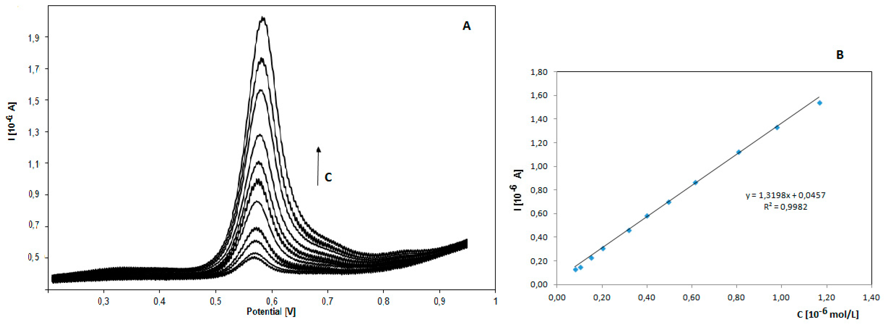
The SWV technique was used to develop the procedure of assaying itraconazole and posaconazole. The study was carried out in experimentally confirmed optimum measurement conditions. In order to determine the relationship between the recorded current and concentration, currents were measured in solutions with growing concentrations of itraconazole. Four series of measurements were carried out, in the concentration range of 7.9 × 10⁻⁸ to 1.2 × 10⁻⁶ mol/L and with potentials from 0.2 V to 0.95 V. The diagram of relationship between current values and the concentration of itraconazole on BDD electrode is presented in Figure 6.
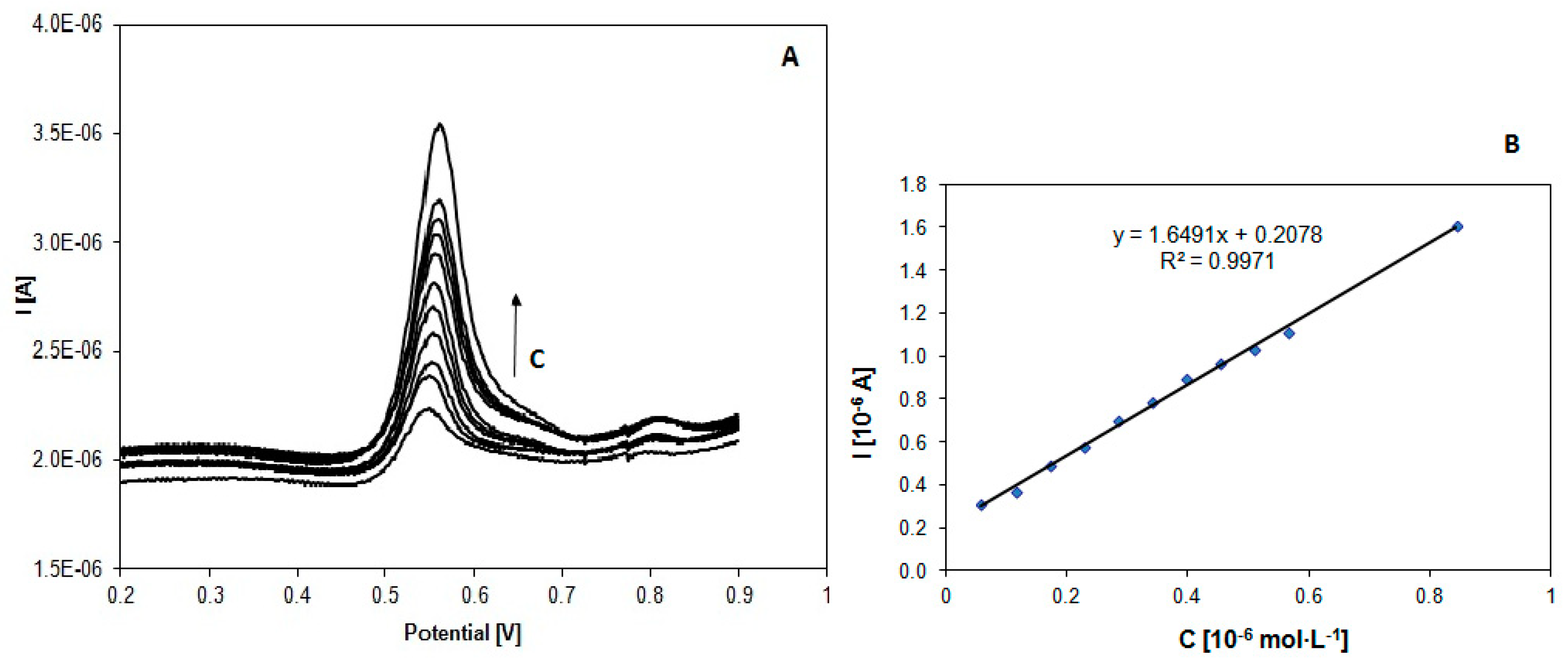
An analogous study of the relationship between the recorded current and the concentration was carried out for posaconazole. The measurements were carried out in the concentration range from 5.7 × 10⁻⁸ to 8.44 × 10⁻⁷ mol·L−1 and the potential range from 0.4 V to 0.75 V. The obtained calibration curve of posaconazole determination and an example series of obtained voltammograms are presented in Figure 7.
The obtained curves are well defined, sharp peaks. Increasing concentration results in higher current values, while the potential does not change. The obtained calibration curves have a coefficient of determination close to one, which proves good correlation of the results. The limit of detection (LOD) and limit of quantification (LOQ) were calculated on the basis of background curves obtained on BDD electrode using the SWV technique. The studied parameters were standard deviation from the obtained background currents (S) and the slope of the calibration curves (m). The following equations were used in the calculations [44]:
LOD = 3.3 S/m, LOQ = 10 S/m
Values characteristic of the calibration curves of itraconazole and posaconazole are presented in Table 1. The obtained data proves the high sensitivity of the proposed procedures.
The developed procedure determination of itraconazole was compared with other methods described in the literature (Table 2). The data presented in Table 2 proves the high sensitivity of the developed procedure against the methods proposed in the literature.
The electrochemical procedure of posaconazole determination developed in the work is proposed in literature for the first time. It is a quick and simple procedure with high sensitivity, comparable with other methods, especially chromatographic ones [49].
3.5. Precision and Selectivity of Itraconazole Using SWV
The precision of the developed procedures was tested. The study of repeatability involved recording voltammetric curves of itraconazole with the concentration of 1.5 × 10⁻⁷ mol/L in the system of several repetitions for the same conditions of analysis. The measurements were performed within one day and over several successive days. The value of absolute standard deviation of measurements obtained within one day was 5.68%, and in the case of five successive days, 2.61%. Analogous studies for posaconazole solution with a concentration of 1.5 × 10⁻⁷ mol/L showed that the value of absolute standard deviation of measurements obtained within one day was 1.73%, and in the case of five successive days, 1.93%. The values of potentials of itraconazole or posaconazole oxidation peak did not change, and the obtained RSD values did not exceed 1%. The obtained results prove the good precision of the developed methods.
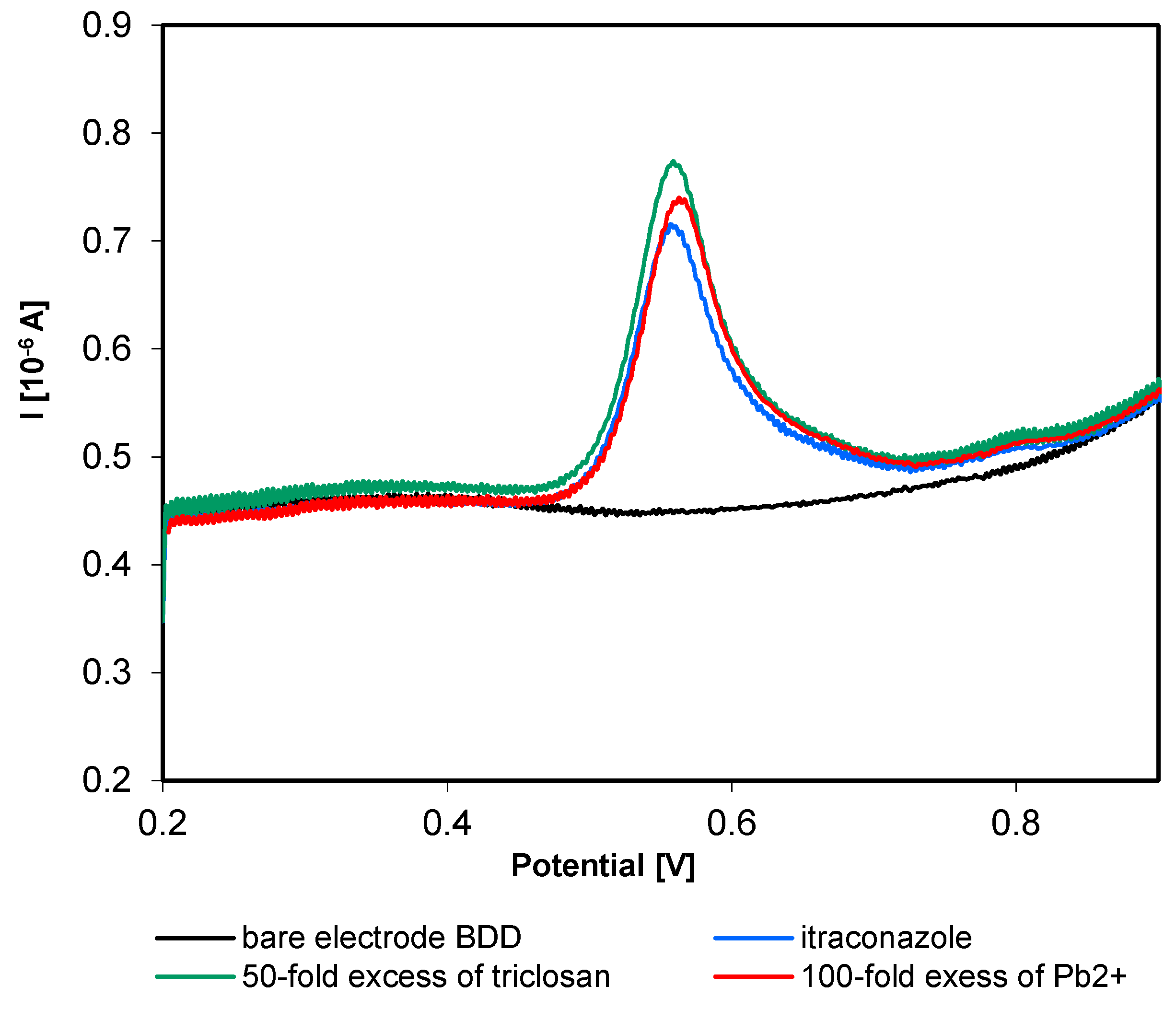
In order to study the selectivity of the developed methods, the impact of electrochemically active substances in the studied range of potentials and substances that can occur in environmental water samples was studied. The analysis involved interferents such as: Na+, K+, Cu2+, Ca2+, Fe2+, Mg2+, Cd2+, Zn2+, Pb2+, Cl−, SO42−, NO3−, Triton X-100, sodium dodecyl sulfate (SDS), tetrabutylamonium bromide, and methylparaben. In addition, experiments were carried out for other fungicides (triclosan) and azole compounds (ketoconazole, clotrimazole, voriconazole). The tests were performed for itraconazole (or posaconazole) solution with the concentration 1.5 × 10⁻⁷ mol/L. Then, 10, 50, 100, 200 and 500-fold excess of the analyzed interferent in relation to the analyte was introduced. The heights of peaks on curves obtained in the presence of potentially interfering substances were compared with the height of peaks obtained for studied biocide alone. It was assumed that the compound does not cause interference if the approximation error of the assay does not exceed ±5%. Based on the obtained results, it was found that itraconazole can be assayed in the presence of inorganic ions with 500-fold excess of the interferent in relation to the concentration of the analyzed compound. Only in the case of Pb2+ ions, it was 100-fold excess in relation to the assayed analyte. Organic compounds such as: Triton X-100 (non-ionic surfactant), SDS (anionic surfactant), tetrabutylamonium bromide (cationic surfactant) and methylparaben allow to assay itraconazole at 100-fold excess of the interferent in relation to the assayed compound. The study of selectivity of itraconazole did not show the presence of new peaks on SWV curves in the presence of interferents. The observed influence was only the increase or decrease in the recorded peaks of the analyte in the presence of interferents (Figure 8).
In the case of posaconazole, the recorded peaks increased at the 100-fold excess of Fe2+ and Cu2+ ions. The other ions did not change the peak heights in the studied range. Just like in the case of itraconazole, the study of surfactants showed interference at an over 100-fold excess, causing the increase in posaconazole oxidation peak. The measurements performed for other fungicides, such as triclosan and ketoconazole, showed the increase in the height of itraconazole and posaconazole oxidation peaks at a 5-fold excess. The study showed that triclosan and ketoconazole have the highest oxidation peaks in solutions with pH = 9. Both posaconazole and itraconazole can be assayed in the presence of azoles such as clotrimazole and voriconazole. Due to the structure of those azoles, different from that of itraconazole or posaconazole, oxidation peaks of these azoles are not observed in the studied potential range. The obtained results prove that the proposed methods display good selectivity.
3.6. Assay of Itraconazole and Posaconazole in the Water Samples
On the basis of the review of methods of assaying itraconazole described in the literature, we can say that biological liquids [45], pharmaceuticals [46] and water samples [11,15] are used as matrices to assay azole fungicides. So as to assess the practical utility of the developed procedure using square wave voltammetry (SWV) and BDD electrode, measurements were carried out in water matrices. The measurements were performed for samples of tap water and river water, which were enriched with known amounts of itraconazole. Methanol solution of itraconazole was added to tap or river water samples to obtain final concentrations: 1.5 × 10⁻⁷ mol/L, 3 × 10⁻⁷ mol/L and 4.5 × 10⁻⁷ mol/L, respectively. Three repetitions were performed for each prepared sample enriched with a specific amount of itraconazole, and then, the mean value was calculated.
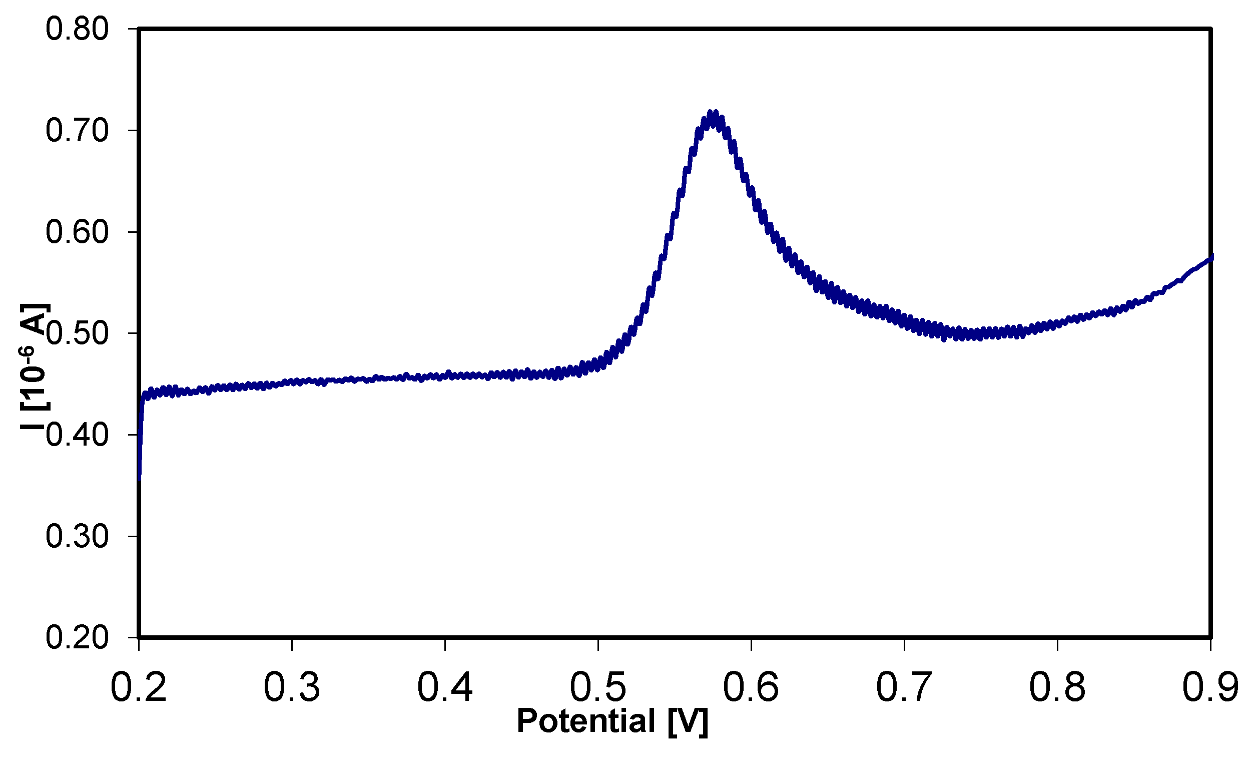
The sample of river water was collected from the Biała river in Białystok, filtrated and kept in a refrigerator before the analysis. The sample of tap water was collected from the waterworks and analyzed without previous preparation. In order to assay itraconazole with the developed procedure in a real sample, the solutions were prepared by adding 10 mL water enriched with itraconazole to 10 mL B-R buffer with pH = 2.87. SWV curves were recorded in the range of potentials from 0.2 V to 0.9 V using previously optimized conditions (amplitude 0.025 V, frequency 100 Hz, step potential 2 mV). Figure 9 shows an example SWV curve of itraconazole assay in a real water sample.
Table 3 shows the results obtained for the assay of itraconazole in water samples. The experiments showed that voltammetric curves obtained for samples of water enriched with itraconazole did not differ in the shape or peak potential values from the peak recorded for the studied analyte in previous stages of the study. The rate of recovery in water samples ranged from 93.5% to 101%, which proves the good precision and accuracy of the developed method.
The analysis of samples of tap water and river water enriched with known amounts of posaconazole was performed in the same way. In order to assay posaconazole in water samples using the developed procedure, we studied solutions with concentrations: 1.5 × 10⁻⁷ mol/L, 3 × 10⁻⁷ mol/L and 4.5 × 10⁻⁷ mol/L. SWV curves were recorded with the use of optimized apparatus parameters and B-R buffer with pH = 4.35. The results of the assay are included in Table 4. The rate of recovery from the measurements in water samples was between 94% and 102.8%. The obtained results confirm the analytical usability of the developed procedures of assaying itraconazole and posaconazole in samples of environmental water.
4. Conclusions
The presented work shows the possibilities of using a new electrochemical electrode material—boron-doped diamond electrode—to assay compounds used as biocides (itraconazole and posaconazole). The electrochemical analysis showed that the selected compounds are electrochemically active and can well be studied and assayed on a BDD electrode. It is so because they undergo the reaction of irreversible electrochemical oxidation, and recorded peaks in the range of potentials from 0.55 V to 0.6 V are well defined and can be used in quantitative analysis. Experiments performed for itraconazole and posaconazole allowed us to develop a fully validated procedures, whose usability were verified for environmental water samples. The developed procedure had good sensitivity in comparison with other electrochemical methods of assaying itraconazole recommended in the literature [46,47]. Only the use of additional adsorption of the analyte on the electrode surface lowers the range of assays, but it makes the analysis longer and more complex [45]. The simplicity, speed and the lack of modification stages of BDD electrode are significant advantages of the proposed method. The electrochemical procedure of posaconazole determination developed in the work is proposed in the literature for the first time. It is a quick and simple procedure with high sensitivity, comparable with other methods, especially chromatographic ones [49]. Whereas, good rate of recovery (93.5–102.8%) proves that the developed procedures can be successfully used to assay itraconazole and posaconazole in water samples.
Author Contributions
Conceptualization, K.M.-Ł.; methodology, K.M.-Ł.; investigation, K.M.-Ł.; writing—original draft preparation, K.M.-Ł., writing—review and editing, K.M.-Ł. and B.S.; supervision, B.S.
Funding
This research received no external funding.
Conflicts of Interest
The authors declare no conflict of interest.
References
- Gros, M.; Petrović, M.; Barcelo, D. Development of multi-residue analytical methodology based on liquid chromatography-tandem mass spectrometry (LC-MS/MS) for screening and trace level determination of pharmaceuticals in surface and wastewaters. Talanta 2006, 70, 678–690. [Google Scholar] [CrossRef]
- Zounkova, R.; Kovalova, L.; Blaha, L.; Dott, W. Ecotoxicity and genotoxicity assessment of cytotoxic antineoplastic drugs and their metabolites. Chemosphere 2010, 81, 253–260. [Google Scholar] [CrossRef]
- Stuart, M.; Lapworth, D.; Crane, E.; Hart, A. Review of risk from potential emerging contaminants in UK groundwater. Sci. Total Environ. 2012, 416, 1–21. [Google Scholar] [CrossRef] [Green Version]
- Hernando, M.D.; Gomez, M.J.; Aguera, A.; Fernandez-Alba, A.R. LC-MS analysis of basic pharmaceuticals (beta-blokers and anti-ulcer agent) in wastewater and surface water. Trends Anal. Chem. 2007, 26, 581–594. [Google Scholar] [CrossRef]
- Hyungkeun, R.; Subramanya, N.; Zhao, F.; Yu, C.; Sandt, J.; Chu, K. Biodegradation potential of wastewater micropollutants by ammonia-oxidizing bacteria. Chemosphere 2009, 77, 1087–1089. [Google Scholar]
- Sirés, I.; Brillas, E. Remediation of water pollution caused by pharmaceutical residues based on electrochemical separation and degradation technologies: A review. Environ. Int. 2012, 40, 212–229. [Google Scholar] [CrossRef] [PubMed]
- European Commission. Directive 98/8/EC of the European Parliament and of the Council (Biocidal Products Directive (BPD) 98/8/EC). Off. J. Eur. Comm. 1998, 41, 123. [Google Scholar]
- Bester, K.; Scholes, L.; Wahlberg, C.; McArdell, C.S. Sources and mass flows of xenobiotics in urban water cycles—An overview on current knowledge and data gaps water. Air Soil Pollut. Focus 2008, 8, 407–4233. [Google Scholar] [CrossRef]
- Chen, Z.; Ying, G.; Lai, H.; Chen, F.; Su, H.; Liu, Y.; Peng, F.; Zhao, J. Determination of bocides in differential environmental matrices by use of ultra-high-performance liquid chromatography-tandem mass spectrometry. Anal. Bioanal. Chem. 2012, 404, 3175–3188. [Google Scholar] [CrossRef]
- Van De Steene, J.C.; Lambert, W.E. Validation of solid-phase extraction and liquid chromatography-electrospray tandem mass spectrometric method for the determination of nine basic pharmaceuticals in wastewater and surface water samples. J. Chromatogr. A 2008, 1182, 153–160. [Google Scholar] [CrossRef] [PubMed]
- Van De Steene, J.C.; Lambert, W.E. Comparison of matrix effects in HPLC-MS/MS and UHPLC-MS/MS analysis of nine basic pharmaceuticals in surface waters. J. Am. Soc. Mass Spectrom. 2008, 19, 713–718. [Google Scholar] [CrossRef]
- Wang, Z.; Zhao, P.; Yu, J.; Jiang, Z.; Guo, X. Experimental and molecular docking study on grapheme/Fe3O4 composites as a sorbent for magnetic solid-phase extraction of seven imidazole antifungals in environmental water samples prior to LC-MS/MS for enantiomeric analysis. Microchem. J. 2018, 140, 222–231. [Google Scholar] [CrossRef]
- Lindberg, R.H.; Fick, J.; Tysklind, M. Screening of antimycotics in Swedish sewage treatment plants—Waters and sludge. Water Res. 2010, 44, 649–657. [Google Scholar] [CrossRef]
- Van De Steene, J.C.; Stove, C.P.; Lambert, W.E. A field study on 8 pharmaceuticals and 1 pesticide in Belgium: Removal rates in waste water treatment plants and occurrence in surface water. Sci. Total Environ. 2010, 408, 3448–3453. [Google Scholar] [CrossRef] [PubMed]
- Castro, G.; Roca, M.; Redriguez, I.; Ramil, M.; Cela, R. Identification and determination of chlorinated azoles in sludge using liquid chromatography quadrupole time-of-flight and triple quadrupole mass spectrometry platforms. J. Chromatogr. A 2016, 1476, 69–76. [Google Scholar] [CrossRef] [PubMed]
- Fatta, D.; Nikolaou, A.; Achilleous, A.; Meric, S. Analytical methods for tracing pharmaceutical residues in water and wastewater. Trends Anal. Chem. 2007, 26, 515–535. [Google Scholar] [CrossRef]
- Renitaa, A.; Senthil, P.; Kumarb, P.; Srinivasb, S.; Priyadharshinib, S.; Karthikab, M. A review on analytical methods and treatment techniques of pharmaceutical wastewater. Desalin. Water Treat. 2017, 87, 160–178. [Google Scholar] [CrossRef] [Green Version]
- Peralta, E.; Natividad, R.; Roa, G.; Marin, R.; Romero, R.; Pavon, T. A comparative study on the electrochemical production of H2O2 between BDD and graphite cathodes. Sustain. Environ. Res. 2013, 23, 259–266. [Google Scholar]
- Panizza, M.; Brillas, E.; Comninellis, C. Application of boron-doped diamond electrodes for wastewater treatment. J. Environ. Eng. Manag. 2008, 18, 139–153. [Google Scholar]
- Einaga, Y. Diamond electrodes for electrochemical analysis. J. Appl. Electrochem. 2010, 40, 1807–1816. [Google Scholar] [CrossRef]
- Luong, J.; Male, K.; Glennon, J. Boron-doped diamond electrode: Synthesis, characterization, functionalization and analytical applications. Analyst 2009, 134, 1965–1979. [Google Scholar] [CrossRef] [PubMed]
- Zhou, Y.; Zhi, J. The application of boron-doped diamond electrode in amperometric biosensors. Talanta 2009, 79, 1189–1196. [Google Scholar] [CrossRef] [PubMed]
- Yang, N.; Yu, S.; Macpherson, J.V.; Einaga, Y.; Zhao, H.; Zhao, G.; Swain, G.M.; Jiang, Y. Conductive diamond: Synthesis, properties, and electrochemical applications. Chem. Soc. Rev. 2019, 48, 157–204. [Google Scholar] [CrossRef] [PubMed]
- Muzyka, K.; Sun, J.; Fereja, T.; Lan, Y.; Zhang, W.; Xu, G. Boron-doped diamond: Current progress and challenges in view of electroanalytical applications. Anal. Methods 2019, 11, 397–414. [Google Scholar] [CrossRef]
- Pecková, K.; Musilová, J.; Barek, J. Boron-doped diamond film electrode—New tool for voltammetric determination of organic substances. Crit. Rev. Anal. Chem. 2009, 39, 148–172. [Google Scholar] [CrossRef]
- Mielech-Łukasiewicz, K.; Rogińska, K. Voltammetric determination of antifungal agents in pharmaceuticals and cosmetics using a boron-doped diamond electrode. Anal. Methods 2014, 6, 7912–7922. [Google Scholar] [CrossRef]
- Mielech-Łukasiewicz, K.; Dąbrowska, A. Comparison of boron-doped diamond and glassy carbon electrodes for determination of terbinafiny in pharmaceuticals using differential pulse and square wave voltammetry. Anal. Lett. 2014, 74, 1697–1711. [Google Scholar] [CrossRef]
- Yardim, Y.; Alpar, N.; Senturk, Z. Voltammetric sensing of triclosan in the presence of cetyltrimethylammonium bromide using a cathodically pretreated boron-doped diamond electrode. Int. J. Environ. Anal. Chem. 2018, 98, 1–16. [Google Scholar] [CrossRef]
- Brocenschi, R.; Rocha-Filho, R.; Biaggio, S.; Bocchi, N. DPV and SWV determination of estrone using a cathodically pretreated boron-doped diamond electrode. Electroanalysis 2014, 26, 1588–1597. [Google Scholar] [CrossRef]
- Sousa, C.P.; Ribeiro, F.W.P.; Oliveira, T.M.; Salazar-Banda, G.R.; de Lima-Neto, P.; Morais, S.; Correia, A.N. Electroanalysis of pharmaceuticals on boron-doped diamond Electrodes: A Review. ChemElectroChem 2019, 6, 2350–2378. [Google Scholar]
- Gayen, P.; Chaplin, B.P. Selective electrochemical detection of ciprofloxacin with a porous nafion/multi-walled carbon nanotube composite film electrode. ACS Appl. Mater. Interfaces 2016, 8, 1615–1626. [Google Scholar] [CrossRef] [PubMed]
- Zhao, Y.; Yuan, F.; Quan, X.; Yu, H.; Chen, S.; Zhao, H.; Liub, Z.; Hilalb, N. An electrochemical sensor for selective determination of sulfamethoxazole in surface water using a molecularly imprinted polymer modified BDD electrode. Anal. Methods 2015, 7, 2693–2698. [Google Scholar] [CrossRef]
- Radicova, M.; Behul, M.; Vojs, M.; Bodor, R.; Vojs Stano, A. Voltammetric determination of erythromycin in water samples using a boron-doped diamond electrode. Phys. Status Solidi B 2015, 252, 2608–2613. [Google Scholar] [CrossRef]
- Calisto, C.; Cervini, P.; Cavalheiro, E.T. Determination of tetracycline in environmental water samples at a graphite-polyurethane composite electrode. J. Braz. Chem. Soc. 2012, 23, 938–943. [Google Scholar] [CrossRef] [Green Version]
- Madej, M.; Kochana, J.; Baś, B. Determination of viloxazine by differential pulse voltammetry with boron-doped diamond electrode. Monatshefte Chem.-Chem. Mon. 2019. [Google Scholar] [CrossRef]
- Ardelean, M.; Manea, F.; Pop, A.; Schoonman, J. Carbon-based electrochemical detection of pharmaceuticals from water. Int. J. Environ. Ecol. Eng. 2016, 10, 1237–1242. [Google Scholar]
- Vojić, M.; Popović, P. Protolytic equilibria in homogeneous and heterogeneous systems of ketoconazole and its direct spectrophotometric determination in tablets. J. Serb. Chem. Soc. 2005, 70, 67–78. [Google Scholar] [CrossRef]
- Popa, O.M.; Diculescu, V.C. Electrochemical and spectrophotometric characterization of proteinkinase inhibitor and anticancer drug danusertib. Electrochim. Acta 2013, 112, 486–492. [Google Scholar] [CrossRef]
- Uslu, B.; Topal, N.; Ozkan, S.A. Electroanalytical investigation and determination of pefloxacin in pharmaceuticals and serum at boron-doped diamond electrodes. Talanta 2008, 74, 1191–1200. [Google Scholar] [CrossRef] [PubMed]
- Sanli, S.; Basaran, F.; Sanli, N.; Akmese, B.; Bulduk, A. Determination of dissociation constants of some antifungal drugs by two different methods at 298K. J. Solut. Chem. 2013, 1976–1987. [Google Scholar] [CrossRef]
- Courney, R.; Wexler, D.; Radwanski, E.; Lim, J.; Laughlin, M. Effect of food on the relative bioavailability of two oral formulations o posaconazole in healty aduits. Br. J. Clin. Pharmacol. 2004, 57, 218–222. [Google Scholar] [CrossRef] [PubMed]
- Alshalalfeh, M.; Sohail, M.; Saleh, T.; Aziz, A. Electrochemical investigation of gold nanoparticle-modified glassy carbon electrode and its application in ketoconazole determination. Aust. J. Chem. 2016, 69, 1314–1320. [Google Scholar] [CrossRef]
- Galus, Z. Fundamentals of Electrochemical Analysis; Ellis Horwood Press: New York, NY, USA, 1994. [Google Scholar]
- Miller, J.C.; Miller, J.N. Statistics for Analytical Chemistry; Ellis Horwood: Chichester, UK; New York, NY, USA, 1988. [Google Scholar]
- Shalaby, A.; Hassan, W.S.; Hendawy, H.A.M.; Ibrahim, A.M. Electrochemical oxidation behavior of itraconazole at different electrodes and its anodic stripping determination in pharmaceuticals and biological fluids. J. Electroanal. Chem. 2016, 763, 51–62. [Google Scholar] [CrossRef]
- Knoth, H.; Knoth, H.; Scriba, G.K.E.; Buettner, B. Electrochemical behavior of the antifungal agents itraconazole, posaconazole and ketoconazole at a glassy carbon electrode. Pharmazie 2015, 70, 374–378. [Google Scholar] [PubMed]
- Knoth, H.; Petry, T.; Gartner, P. Differential pulse polarographic investigation of the antifungal drugs itraconazole, ketoconazole, fluconazole and voriconazole using a dropping mercury electrode. Pharmazie 2012, 67, 987–990. [Google Scholar]
- Sultan, M.A.; Attia, A.K.; El-Alamin, M.M.A.; Atia, M.A. The novel use of multwalled carbon nanotubes-based sensors for voltammetric determination of itraconazole: Application to pharmaceutical dosage form and biological samples through spiked urine sample. World J. Pharm. Sci. 2016, 5, 93–108. [Google Scholar]
- Reddy, T.M.; Tama, C.; Hayes, R.N. A dried blond spots technique based LC-MS/MS method for the analysis of posaconazole in human whole blood samples. J. Chromatogr. B 2011, 879, 3626–3638. [Google Scholar] [CrossRef]
Figure 1.
Structure of studied biocides: (A) itraconazole, (B) posaconazole.

Figure 2.
Cyclic voltammograms of 2.7 × 10⁻⁵ mol·L−1 posaconazole in Britton-Robinson (B-R) buffer in the pH 1.8–11.2 range; v = 100 mV·s−1.
Figure 2.
Cyclic voltammograms of 2.7 × 10⁻⁵ mol·L−1 posaconazole in Britton-Robinson (B-R) buffer in the pH 1.8–11.2 range; v = 100 mV·s−1.

Figure 3.
Cyclic voltammograms of 2.7 × 10⁻⁵ mol·L−1 itraconazole in B-R buffer in the pH 1.8–11.2 range; v = 100 mV·s−1.
Figure 3.
Cyclic voltammograms of 2.7 × 10⁻⁵ mol·L−1 itraconazole in B-R buffer in the pH 1.8–11.2 range; v = 100 mV·s−1.

Figure 4.
Effect of pH on selected biocides: (A) anodic peak current and (B) anodic peak potential.


Figure 5.
Cyclic voltammograms of 2.44 × 10⁻⁵ mol·L−1 itraconazole in B-R buffer pH 2.87 at different scan influence of interferents rates (A); the dependence of Ip on the square root of scan rate (B); and the plot for dependence of log Ip on log of scan rate (C).
Figure 5.
Cyclic voltammograms of 2.44 × 10⁻⁵ mol·L−1 itraconazole in B-R buffer pH 2.87 at different scan influence of interferents rates (A); the dependence of Ip on the square root of scan rate (B); and the plot for dependence of log Ip on log of scan rate (C).

Figure 6.
Square wave voltammetry (SWV) voltammograms of different concentrations of itraconazole in B-R buffer pH 2.87 (range of concentration 7.9 × 10⁻⁸ to 1.2 × 10⁻⁶ mol·L−1) (A); Calibration curves of itraconazole using SWV methods, on the boron-doped diamond (BDD) electrode (B).
Figure 6.
Square wave voltammetry (SWV) voltammograms of different concentrations of itraconazole in B-R buffer pH 2.87 (range of concentration 7.9 × 10⁻⁸ to 1.2 × 10⁻⁶ mol·L−1) (A); Calibration curves of itraconazole using SWV methods, on the boron-doped diamond (BDD) electrode (B).

Figure 7.
SWV voltammograms of different concentrations of posaconazole in B-R buffer pH 4.35 (range of concentration 5.7 × 10⁻⁸–8.44 × 10⁻⁷ mol·L−1) (A); Calibration curves of posaconazole using SWV methods, on the BDD electrode (B).
Figure 7.
SWV voltammograms of different concentrations of posaconazole in B-R buffer pH 4.35 (range of concentration 5.7 × 10⁻⁸–8.44 × 10⁻⁷ mol·L−1) (A); Calibration curves of posaconazole using SWV methods, on the BDD electrode (B).

Figure 8.
SWV voltammograms of itraconazole in B-R buffer pH 2.87 in the presents of selected interferents.
Figure 8.
SWV voltammograms of itraconazole in B-R buffer pH 2.87 in the presents of selected interferents.

Figure 9.
Square wave voltammogram of itraconazole in water samples.


Table 1.
Quantitative determination of itraconazole and posaconazole on the BDD electrode using the SWV method.
Table 1.
Quantitative determination of itraconazole and posaconazole on the BDD electrode using the SWV method.
| Studied Substance | Itraconazole | Posaconazole |
|---|---|---|
| Peak potential/V vs. SCE | 0.59 | 0.55 |
| Peak width half/mV | 0.06 | 0.08 |
| Linearity range/mol·L−1 | 7.9 × 10⁻⁸–1.2 × 10⁻⁶ | 5.7 × 10⁻⁸–8.44 × 10⁻⁷ |
| Slope/µA·L/mol | 1.32×10⁶ | 1.65 × 10⁶ |
| Intercept/µA | 0.046 | 0.2 |
| Correlation coefficient | 0.9982 | 0.9971 |
| LOQ/mol·L−1 | 5.43 × 10⁻⁸ | 2.36 × 10⁻⁸ |
| LOD/mol·L−1 | 1.79 × 10⁻⁸ | 7.78 × 10⁻⁹ |
| Repeatability of Ip/RSD% | 5.68 | 1.73 |
| Reproducibility of Ip/RSD% | 2.61 | 1.93 |
Note: SCE: saturated calomel electrode; LOQ: limit of quantification; LOD: limit of detection.
Table 2.
Comparison of linear range and detection limits for itraconazole to different methods.
| Linear Range (mol·L−1) | Detection Limit (mol·L−1) | Method | Electrode | Ref. |
|---|---|---|---|---|
| 2.2 × 10⁻⁸–2.9 × 10⁻⁷ | 1.9 × 10⁻⁸ | UTG | [45] | |
| 1.5 × 10⁻⁸–2.3 × 10⁻⁷ | 1.2 × 10⁻⁸ | AS-SWV | PG | |
| 1.5 × 10⁻⁸–1.5 × 10⁻⁷ | 8.5 × 10⁻⁹ | CP | ||
| 2.2 × 10⁻⁸–2.5 × 10⁻⁷ | 1.5 × 10⁻⁸ | UTG | ||
| 4.5 × 10⁻⁸–2.3 × 10⁻⁷ | 1.2 × 10⁻⁸ | AS-DPV | PG | |
| 1.5 × 10⁻⁸–1.5 × 10⁻⁷ | 1.1 × 10⁻⁸ | CP | ||
| 2.8 × 10⁻⁵–1.4 × 10⁻⁴ | - | CV | GC | [46] |
| 5.0 × 10⁻⁷–5.0 × 10⁻⁶ | - | DPV | Hg | [47] |
| 2.19 × 10⁻⁶–6.33 × 10⁻⁵ | 7.27 × 10⁻⁷ | CV | MWCNT/CP | [48] |
| 7.9 × 10⁻⁸–1.2 × 10⁻⁶ | 1.79 × 10⁻⁸ | SWV | BDD | This work |
Note: UTG ultra-trace graphite, PG pencil graphite, GC glassy carbon, CP carbon paste, BDD boron doped diamond, MWCNT multi-walled carbon nanotube, CV cyclic voltammetry, DPV differential pulse voltammetry, SWV square wave voltammetry, AS-SWV anodic stripping square wave voltammetry, AS-DPV anodic stripping differential pulse voltammetry.
Table 3.
Results of itraconazole determination in spiked water samples.
| Sample | Amount Added/×10−7 mol/L | Amount Received a/×10−7 mol/L | Recovery a/% | RSD/% |
|---|---|---|---|---|
| Biała water | 1.500 | 1.437 ± 0.069 | 95.8 ± 4.5 | 4.8 |
| 3.000 | 2.882 ± 0.107 | 96.1 ± 3.6 | 3.7 | |
| 4.500 | 4.545 ± 0.102 | 101 ± 2.2 | 2.2 | |
| Tap water | 1.500 | 1.403 ± 0.059 | 93.5 ± 3.9 | 4.2 |
| 3.000 | 2.919 ± 0.056 | 97.3 ± 1.9 | 1.9 | |
| 4.500 | 4.350 ± 0.145 | 96.7 ± 3.2 | 3.3 |
a Mean value (n = 3).
Table 4.
Results of posaconazole determination in spiked water samples.
| Sample | Amount Added/×10−7 mol/L | Amount Received a/×10−7 mol/L | Recovery a/% | RSD/% |
|---|---|---|---|---|
| Biała water | 1.500 | 1.438 ± 0.052 | 95.8 ± 3.4 | 3.6 |
| 3.000 | 2.821 ± 0.079 | 94.0 ± 2.6 | 2.8 | |
| 4.500 | 4.399 ± 0.115 | 97.7 ± 2.5 | 2.6 | |
| Tap water | 1.500 | 1.447 ± 0.023 | 96.5 ± 1.6 | 1.6 |
| 3.000 | 2.886 ± 0.102 | 96.2 ± 3.4 | 3.5 | |
| 4.500 | 4.627 ± 0.055 | 102.8 ± 1.2 | 1.2 |
a Mean value (n = 3).
© 2019 by the authors. Licensee MDPI, Basel, Switzerland. This article is an open access article distributed under the terms and conditions of the Creative Commons Attribution (CC BY) license (http://creativecommons.org/licenses/by/4.0/).
Share and Cite
MDPI and ACS Style
Mielech-Łukasiewicz, K.; Starczewska, B. The Use of Boron-Doped Diamond Electrode for the Determination of Selected Biocides in Water Samples. Water 2019, 11, 1595. https://doi.org/10.3390/w11081595
AMA Style
Mielech-Łukasiewicz K, Starczewska B. The Use of Boron-Doped Diamond Electrode for the Determination of Selected Biocides in Water Samples. Water. 2019; 11(8):1595. https://doi.org/10.3390/w11081595
Chicago/Turabian StyleMielech-Łukasiewicz, Katarzyna, and Barbara Starczewska. 2019. "The Use of Boron-Doped Diamond Electrode for the Determination of Selected Biocides in Water Samples" Water 11, no. 8: 1595. https://doi.org/10.3390/w11081595
Note that from the first issue of 2016, this journal uses article numbers instead of page numbers. See further details here.