Removal of Pb(II) by Pellicle-Like Biofilm-Producing Methylobacterium hispanicum EM2 Strain from Aqueous Media


1
School of Environmental Engineering, University of Seoul, Seoul 02504, Korea
2
World Institute of Kimchi, Gwangju 61755, Korea
*
Authors to whom correspondence should be addressed.
Water 2019, 11(10), 2081; https://doi.org/10.3390/w11102081
Submission received: 26 August 2019
/
Revised: 30 September 2019
/
Accepted: 3 October 2019
/
Published: 5 October 2019
(This article belongs to the Special Issue Technologies Developing in Heavy Metals' Removal from Water)
Abstract
:As concerns are increasing about drinking water contamination with heavy metals, we investigated the possible use of a pellicle (floating biofilm)-like biofilm-producing microorganism as a biosorbent for the treatment of Pb(II) in aqueous solutions. The bacterial pellicle-producing Methylobacterium hispanicum EM2 strain (EM2) was newly isolated from mine tailing soil, and we investigated its use as a biosorbent for treating a Pb(II)-contaminated aqueous solution. The EM2 strain was strongly resistant to Pb(II) up to a concentration of 800 mg/L, and achieved remarkable adsorption performance (adsorption rate and maximum adsorption capacity of 96% ± 3.2% and 79.84 mg/g, respectively) under optimal conditions (pH, biomass content, contact time, and initial Pb(II) concentration of 7.1 g/L, 60 min, and 10 mg/L, respectively). The adsorption of Pb(II) was characterized by scanning electron microscopy-energy dispersive x-ray spectroscopy and Fourier-transform infrared analysis. The equilibrium data matched the Freundlich isotherm model well, indicating the occurrence of multilayer adsorption of Pb(II) onto the heterogeneous surface of the EM2 strain, which was also consistent with the pseudo-second-order kinetic model (R2 = 0.98). The high Pb(II) removal efficiency was also confirmed by conducting an adsorption experiment using Pb(II)-contaminated industrial wastewater.
1. Introduction
Due to the increasing concern about the public health and environmental problems caused by Pb(II) contamination, developing highly efficient and stable treatment methods is necessary. Although Pb(II) is one of the most widely used nonferrous metals that is necessary for manufacturing daily necessities, it has profound negative effects on living organisms [1]. Many toxicological studies have reported abnormal symptoms and diseases caused by the accumulation of Pb(II) in human tissues. For examples, hematopoiesis (anemia) and malfunctions of the nervous, digestive, and cardiovascular systems have been triggered by exposure to Pb(II), even at low concentrations. Pb(II) can enter the human body through ingestion, inhalation, and adsorption, which can cause other severe effects, such as permanent damage to cognition and the reproductive system, and neurodegenerative diseases [2,3]. Thus, the Agency for Toxic Substance and Disease Registry (ATSDR) and the World Health Organization (WHO) have both designated Pb(II) as an extremely hazardous substance, and have withdrawn its provisional value.
To date, much effort has been dedicated to developing an efficient treatment method for removing Pb(II), such as ion exchange, solvent extraction, adsorption, membrane filtration, precipitation, and reverse osmosis [4,5,6,7,8,9]. However, several hindrances, such as (1) difficulties in adsorbent preparation, (2) the risk of secondary contamination, and (3) the requirement for additional steps, such as resin regeneration or replacement, should be overcome prior to practical application. Therefore, collective approaches that combine chemistry and biology to overcome the drawbacks to the conventional process currently in use must be implemented to develop an efficient Pb(II) treatment method. Of the various potential methods, biosorption, which uses inherent biological features that interact with chemicals, is one of the most promising methods for treating toxic heavy metals as it is ecologically sustainable, inexpensive, and the operation parameters can be easily controlled.
Bacterial biofilms are composed of polysaccharides, extracellular DNA, lipids, and other biopolymers [10], and therefore play an important role in the cellular adhesion, attachment, and diffusion of a material. In most cases, biofilm-producing microorganisms can quickly and firmly attach to a target surface via electrostatic attraction (such as van der Waals forces) between the negatively charged biofilms [11] and a positively charged target surface. Most of the heavy metals that are likely to pollute drinking water are positively charged. Thus, the positively charged toxic heavy metals can adhere to the negatively charged biofilms through electrostatic attraction. Therefore, biofilms extracted from various microorganisms in vitro have been examined as potential biosorbents [12,13,14]. However, the adsorption of Pb(II) by pellicle-like biofilm-producing microorganisms has not yet been reported.
As a biosorbent, biofilm-producing microorganisms have a considerable advantage over biofilms extracted in vitro for application in biosorption as they have substantial quantities of anionic functional groups, such as carboxyl-, hydroxyl-, phosphoryl, and amino groups, on their cellular membranes. The metal-binding forces of biofilm-producing microorganisms were found to be 20-times higher, and the interactions with and immobilization of cationic metal ions are more efficient [15,16]. A floating biofilm (pellicle) could be more effective for adsorbing metal ions given their higher bio-volume, roughness, and thickness than submerged biofilms [10,17].
Together, these observations led us to investigate the potential of pellicle-like biofilm-producing microorganism for the removal Pb(II) by electrostatic attraction. This is the first proof-of-concept study to propose the potential of a pellicle-like biofilm-producing microorganism for the treatment of heavy metals from aqueous media.
2. Materials and Methods
2.1. Soil Sampling and Identification of Methylobacterium hispanicum EM2 Strain
A soil sample was collected from the mine tailings in Cheonan, Republic of Korea (36°54′10.19′′ N, 127°15′17.72′′ E). One gram of the soil sample was suspended in sterile saline (0.85%, w/v NaCl) and serially diluted. The dilutions were spread onto tryptone, glucose, and yeast (TGY) agar plates (0.5% tryptone, 0.1% glucose, and 0.3% yeast extract). The isolated bacterial strains were routinely cultured in the TGY medium at 30 °C, with or without agar.
The full length of the 16S rRNA gene was determined using SeqBuilder software (DNASTAR Inc., Madison, WI, USA) and compared with GenBank entries using the Basic Local Alignment Tool (BLAST), a program developed by the National Center for Biotechnology Information (NCBI, http://www.ncbi.nlm.nih.gov). Phylogenetic analysis was conducted following previously described methods [18]. Multiple alignments and phylogenetic trees were reconstructed using the Mega 7.0 program [19] with the neighbor-joining method [20] and Kimura two-parameter model [21] with bootstrap values based on 1000 replications [22].
The isolates were Gram-stained following previously described methods [23]. The phenotypic characteristics of the isolates were determined using an API20NE kit (BioMérieuxTM, Marcy-l'Etoile, France) following the manufacturer’s instructions. The growth of the isolates at different temperatures, NaCl concentrations, and pH values were evaluated on TGY agar at 30 °C for three days. A motility test was conducted on a semi-solid medium.
2.2. Effect of Pb(II) on Bacterial Growth
Cobalt(II) chloride hexahydrate (CoCl2·6H2O), zinc sulfate heptahydrate (ZnSO4·7H2O), nickel(II) chloride hexahydrate (NiCl2·6H2O), copper sulfate pentahydrate (CuSO4·5H2O), lead(II) chloride (PbCl2), and cadmium chloride hydrate (CdCl2·H2O) (Sigma Aldrich, St. Louis, MO, USA) were used to determine the minimal inhibitory concentrations (MICs) of heavy metals following the solid agar method. To prepare the samples, a fresh colony was inoculated at 30 °C in 3 mL of TGY broth. After cultivation, the cells were serially diluted with 0.85% saline. Ten microliters of the dilutes were dropped onto TGY agar plates supplemented with 1–10 mM of the metal solutions and incubated at 30 °C until colonies were visible. To investigate the effect of microbial activity on Pb(II) ions, overnight cell cultures were diluted 1:100 in fresh TGY broth supplemented with different concentrations of PbCl2 (0, 50, 100, 250, 500, and 750 mg/L) and then incubated at 30 °C for 72 h with agitation at 200 rpm. Cell growth was monitored by measuring the optical density at 600 nm (OD600) using an Epoch microplate spectrophotometer (Biotek, Winooski, VT, USA).
2.3. Biosorption Profile
The overnight culture was subcultured (1:100, v/v) in 500 mL of the same liquid medium until the stationary phase was reached. The cultured cells were harvested then rinsed twice with deionized water to remove any residual media components. The sample was freeze-dried and stored at −80 °C before use in the biosorption experiments.
The effect of several parameters, such as pH, contact time, biomass dosage, and initial Pb(II) concentration, on the biosorption efficiency of the biomass was evaluated. To determine the optimum pH value, biosorption experiments were conducted in an Erlenmeyer flask containing 10 mL of a Pb(II) solution (initial concentration of 10 mg/L) for 60 min at 30 °C with agitation at 150 rpm. The initial pH of the metal solution was adjusted using 0.1 M HCl or NaOH solutions. The residual Pb(II) in the supernatant was measured by inductively coupled plasma-optical emission spectroscopy (ICP-OES, iCAPTM 7000 series, Thermo Scientific™, Waltham, MA, USA). All biosorption assays were conducted in triplicate, and the mean values were calculated.
The biosorption capacity and biosorption rate of metal ions were determined using Equations (1) and (2), respectively [24]:
where qe (mg/g) is the equilibrium metal ion concentration on the biosorbent; C0 and Ce (mg/L) are the initial and final concentrations of metal ions in the aqueous solution, respectively; and X is the biomass concentration (g dry cell/L).
2.4. Study of Biosorption and Kinetic Isotherms
The equilibrium isotherms were determined using the Langmuir and Freundlich isotherm models, which are widely used to describe the adsorption mechanism between an adsorbent and absorbate. The Langmuir adsorption isotherm assumes that the adsorbent has a monolayer and a homogeneous surface. However, the Freundlich adsorption isotherm assumes that the adsorbent has a heterogeneous surface with non-uniform adsorption heat and affinity distributions [25]. The Langmuir isotherm model is represented by [26]:
where Ceq (mg/L) is the concentration of metal ions in the aqueous solution at equilibrium, and Qmax (mg/g) and KL (L/mg) are the maximum adsorption capacity and Langmuir adsorption constant related to the affinity of the binding sites, respectively.
The Freundlich model is expressed as [27]:
where KF (L/g) and n are the Freundlich constants correlated with the binding capacity and the affinity of the adsorbent, respectively.
Adsorption kinetics are used to predict the adsorption capacity and reaction rate of the adsorbent [28]. Both pseudo-first-order and pseudo-second-order equations are used to study the kinetics of metal biosorption, as shown in Equations (5) and (6), respectively:
where qt is the adsorbed metal ion concentration (mg/g) at time t (min), qe is the concentration of adsorbed metal ions (mg/g) at equilibrium, and K1 and K2 are the pseudo-first-order (L/min) and pseudo-second-order (g/mg/min) adsorption rate constants, respectively.
2.5. Scanning Electron Microscope Energy Dispersive X-ray Spectroscopy Analysis
The cell morphology and presence of lead on the surfaces were observed using a field emission scanning electron microscope (Inspect F50, FEI, Hillsboro, OR, USA). The elemental composition was analyzed using a scanning electron microscope-energy dispersive X-ray (SEM-EDX) spectrophotometer (EDAX Apollo XL, AMETEK, Mahwah, NJ, USA) with accelerating voltages up to 20 kV. The EDX spectra were recorded in the area scan mode by focusing the electron beam onto a region of the surface of the sample.
2.6. Fourier-Transform Infrared Spectroscopy Analysis
The functional groups that interacted with metal ions on the surfaces of the cells were analyzed using Fourier-transform infrared (FTIR) spectroscopy. The FTIR spectra of the cells before and after treatment with a 10 mg/L Pb(II) solution were recorded using a Nicolet iS10 (ThermoScientific, Waltham, MA, USA) spectrophotometer. The infrared spectra of the samples were captured at a wavenumber range of 500 to 4000 cm−1 and resolution of 1 cm−1.
2.7. Pb(II) Removal from Sewage Water
Sewage water was collected from the sedimentation basin area of the Jungnang sewage treatment plant in Seoul, Republic of Korea. The collected sewage water was filtered through a 1.2 μm Whatman glass microfiber filter (Sigma-Aldrich, St. Louis, MO, USA) and autoclaved at 121 °C for 30 min. The Pb(II) stock solution was added to the sewage water to achieve a final concentration of 10 mg/L. To determine the Pb(II) removal efficiency, 1 g/L of biomass was added to 50 mL of the Pb(II)-contaminated sewage water and incubated at 30 °C for 1 h at 200 rpm.
3. Results and Discussion
3.1. Identification of the Pellicle-Like Biofilm-Producing M. hispanicum EM2 Strain
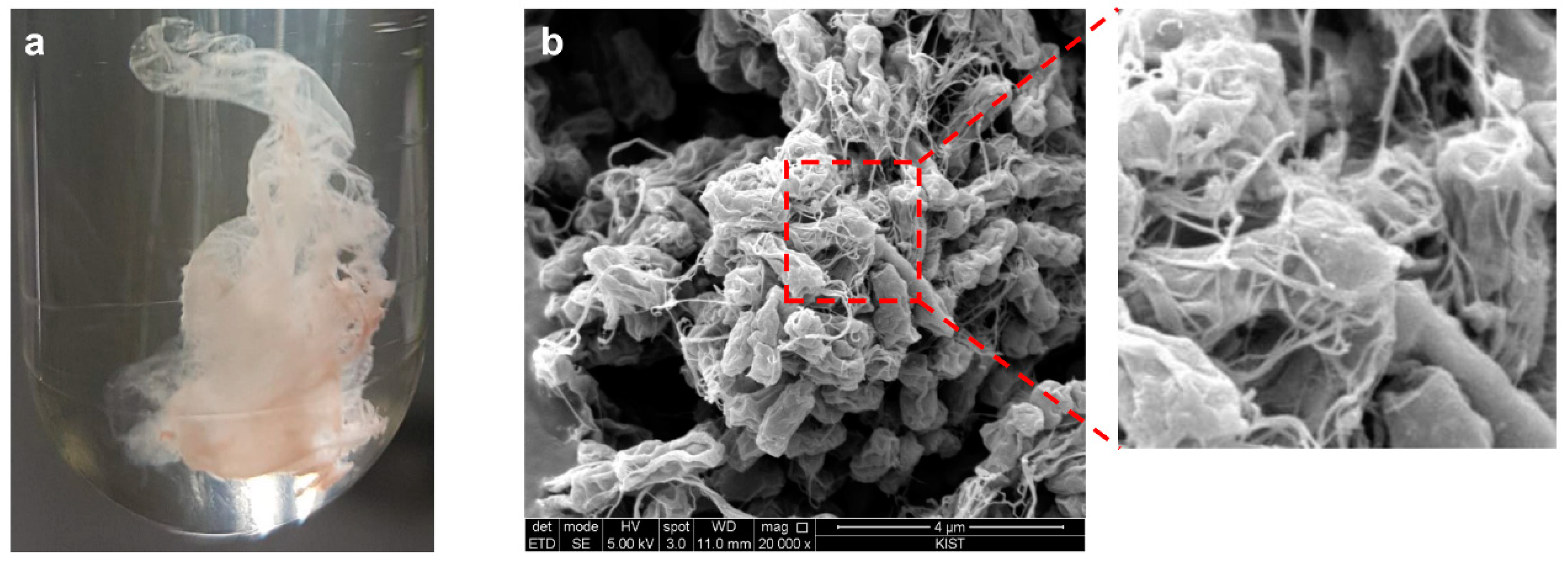
The pellicle-like biofilm-producing Methylobacterium hispanicum EM2 strain (EM2) was newly isolated from mining tail soil (Figure 1). As shown in Table S1, the EM2 strain bacteria were Gram-negative, motile, aerobic, rod-shaped, and red in color, and grew best at pH 7.0 and 30 °C in a TGY culture medium. An analytical profile index (API) test, which is widely used to identify the biochemical properties of a microorganism, was conducted, and this bacterium was scored positive for enzyme activity, glucose fermentation, indole production, and nitrate reduction. This strain could use maleic acid and trisodium citrate as sole carbon sources. The 16S rRNA gene sequence analysis revealed that the EM2 strain is closely related to the Methylobacterium genus, a methylotrophic bacteria, and exhibited high similarity (98.7%) to the Methylobacterium hispanicum GP34T strain [29] (Figure S1).
3.2. Resistance to Pb(II)
In the development of a biosorbent for the removal of toxic heavy metals, the degree of tolerance to the toxic heavy metal is an important indicator when determining whether the biosorbent can be used to purify a contaminated area [30]. Thus, the minimum inhibitory concentration (MIC) was tested to determine the strain’s degree of resistance to various toxic heavy metals, including Co(II), Zn(II), Cu(II), Pb(II), Ni(II), and Cd(II), which all cause environmental pollution. As shown in Figure S2a, the EM2 strain exhibited strong resistance to Pb(II) up to a concentration of 834 mg/L, which is the highest value reported in the biological treatment of Pb(II) to date [31,32,33], but exhibited relatively low tolerance to the other heavy metals (Cd(II), 18 mg/L; Co(II), 130 mg/L; Ni(II), 130 mg/L; Zn(II), 252 mg/L; Cu(II), 269 mg/L). Although cell growth tended to decrease slightly as the concentration of Pb(II) increased, the EM2 strain maintained cellular growth, even at a Pb(II) concentration of 500 mg/L. Thus, the EM2 strain can be considered a biosorbent for the removal of high concentrations of Pb(II) contaminant (Figure S2b).
3.3. Optimization and Isotherm and Kinetic Modeling of the Biosorption of Pb(II)
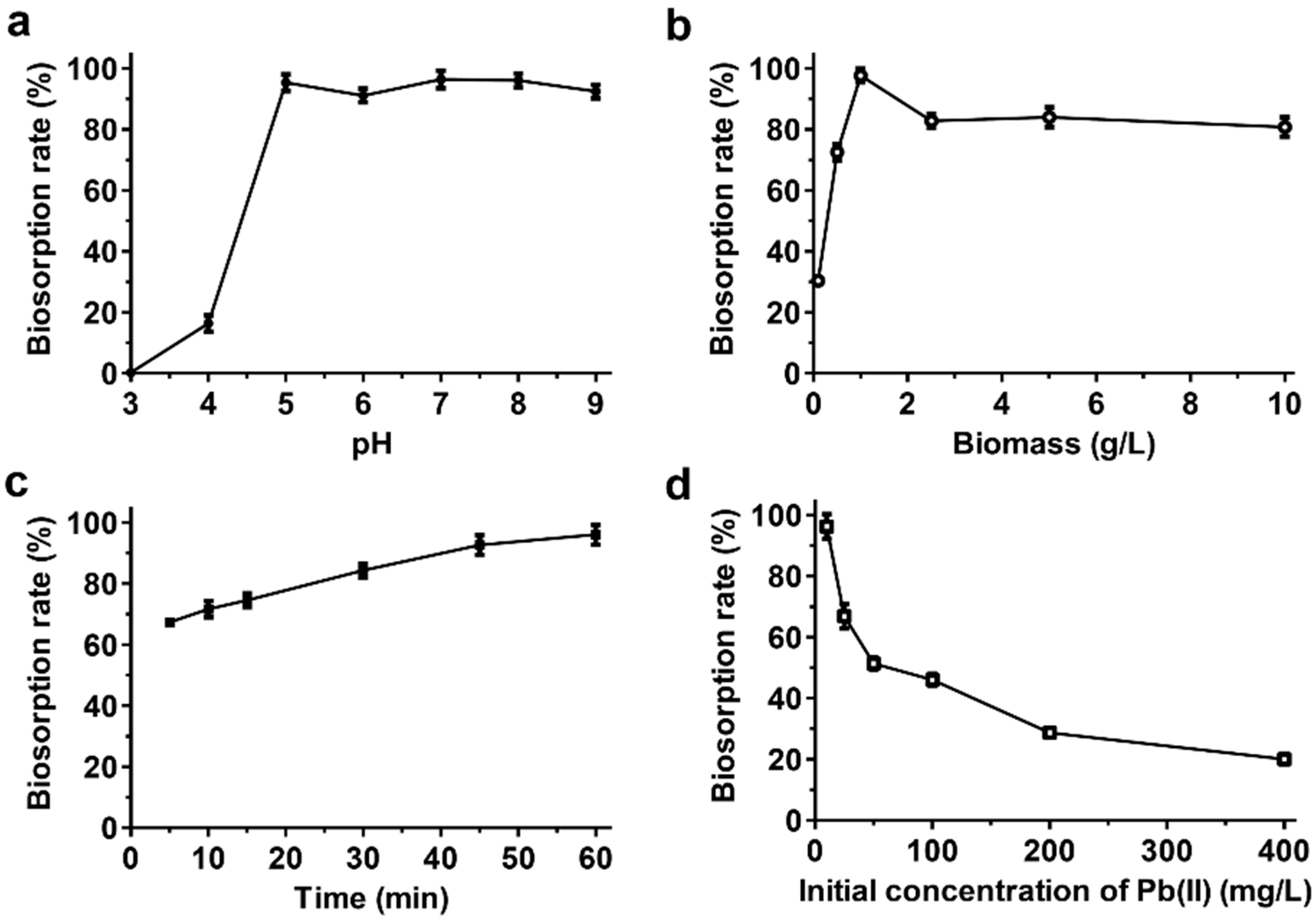
Before investigating the removal capacity of the EM2 strain, various external parameters that are known affect biosorption, such as the pH, amount of biomass, contact time, and initial Pb(II) concentration, were optimized [34]. As shown in Figure 2, the maximum removal efficiency of the EM2 strain was achieved at pH 7 (96.36% ± 2.8%), biomass content of 1 g/L (97.6% ± 2.1%), contact time of 60 min (96% ± 3.2%), and initial Pb(II) concentration of 10 mg/L (96.2% ± 4%).
To understand the process of Pb(II) biosorption by the EM2 strain, the adsorption data were further analyzed using Langmuir and Freundlich models (Figure S3). The biosorption process of the EM2 strain better matched the Freundlich biosorption isotherm model (R2 = 0.976) than the Langmuir model (R2 = 0.962), according to the higher correlation coefficient value (Table 1). Thus, the biosorption of Pb(II) by the EM2 strain could be explained as heterogeneous surface-mediated multiple-layer adsorption caused by bacterial biofilm, as reported in previous studies [35,36]. A kinetics study was also conducted to determine the contact time necessary to reach equilibrium status and optimize conditions for the treatment of Pb(II)-contaminated environments [37]. The pseudo-first-order [38] and pseudo-second-order [39] kinetic models were used to analyze the kinetics of Pb(II) biosorption. The parameters of the kinetic models were calculated by conducting linear regression analysis on the results of independent biosorption experiments conducted under optimal conditions (Figure S3). The Pb(II) adsorption kinetics fitted the pseudo-second-order model well as the experimental value (9.32 mg/g) was similar to the value calculated by the pseudo-second-order model (9.07 mg/g; Table 1). The R2 value of the pseudo-second-order kinetic model for Pb(II) biosorption (0.98) was higher than that of the pseudo-first-order kinetic model (0.97). Thus, we predicted that adsorption was achieved through chemisorption [40].
3.4. Removal of Pb(II) Using the EM2 Strain
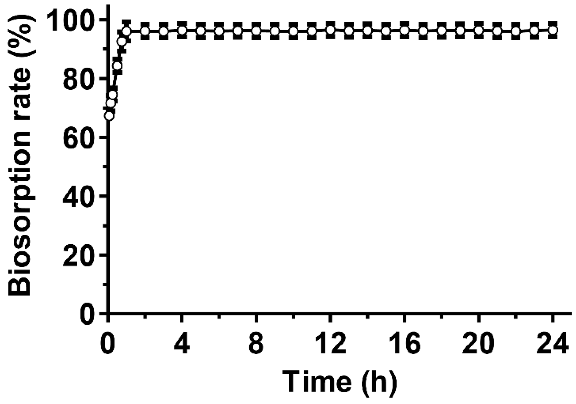
Next, we investigated the Pb(II) removal capacity of the EM2 strain by biosorption in aqueous media. As shown in Figure 3, over 96% of the Pb(II) was successfully removed within one hour, and the maximum removal efficiency remained stable for 24 h. The maximum Pb(II) adsorption capacity obtained was 79.84 ± 5.7 mg/g under optimal conditions, which is higher than that reported in previous biosorption studies (Table 2).
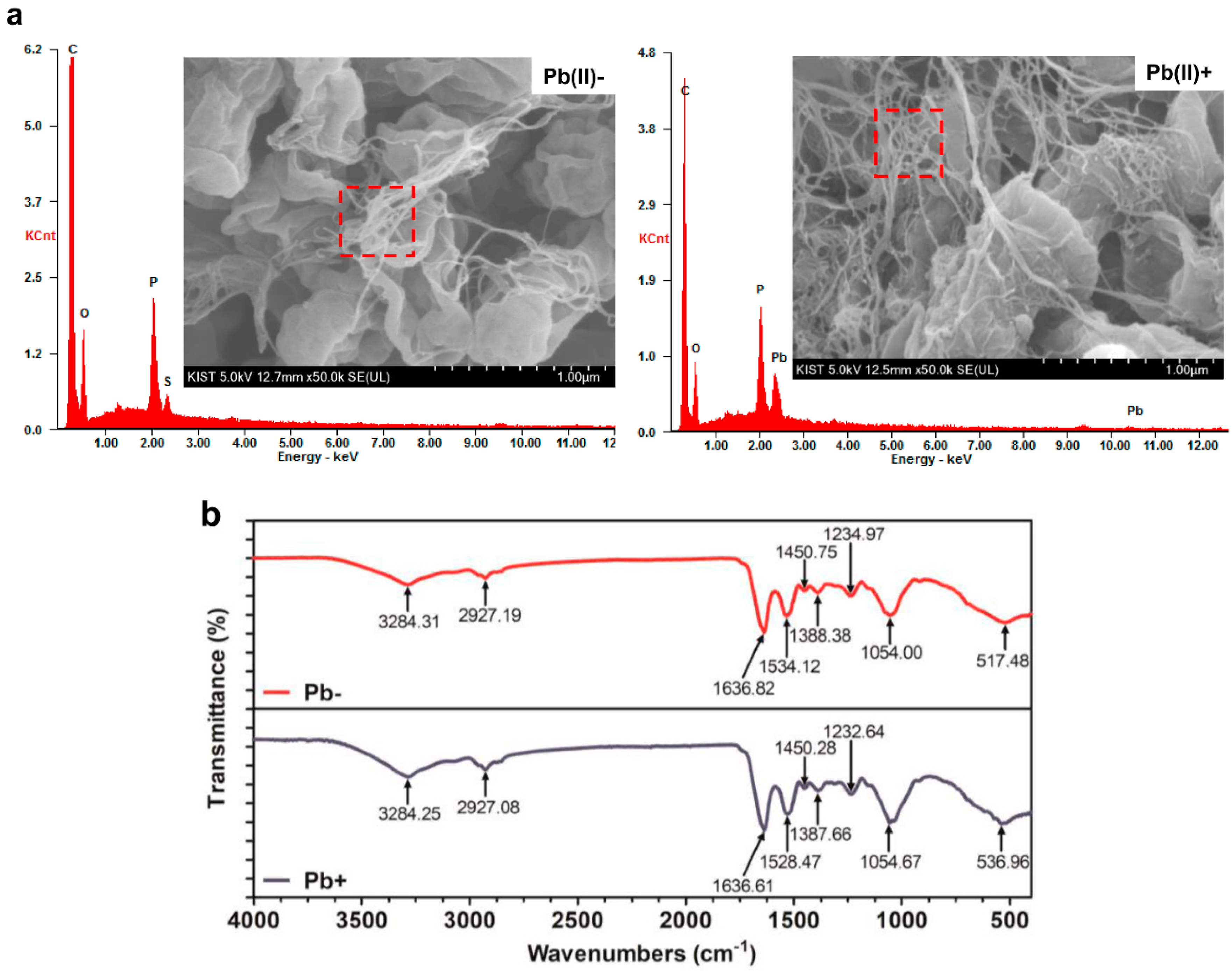
The peaks corresponding to Pb(II) in the EDX spectrum indicated that Pb(II) was selectively adsorbed by the pellicle-like biofilm matrix produced by the EM2 strain (Figure 4a). The peak corresponding to sulfur disappeared after the biosorption of Pb(II), indicating that sulfide groups are involved in the adsorption of Pb(II). In the FTIR spectrum (Figure 4b), the peak at 517.48 cm−1, corresponding to C–N–S scissoring in polypeptides [49,50], shifted to 536.96 cm−1, indicating that the sulfide group is involved in the biosorption of Pb(II), as confirmed by the EDX analysis. The peaks at 1534.12 and 1234.97 cm−1, corresponding to C–N stretching and N–H bending in amide II [42] and phosphate [51], respectively, shifted to 1528 and 1235.64 cm−1, respectively. Based on these results, both amide II and the phosphate group play important roles in the biosorption of Pb(II).
Morphological changes were not observed after treating Pb(II); however, previous studies reported cytomorphological changes after exposure to toxic heavy metals [43,52]. We could assume that most of the heavy metals were adsorbed on the biofilm rather than the cell surface of the EM2 strain owing to the strong electrostatic attraction of the pellicle-like biofilm. Strong electrostatic biofilm forces were also observed in a previous study that explored the components of the M. organophilum biofilm, which has the same lineage as the EM2 strain. The biofilm was mainly composed of negatively charged uronic, pyruvic, and acetic acid, suggesting that it could strongly adsorb positively charged metal ions, such as Cu(II) and Pb(II) [12]. The amount of biofilm increased when the EM2 strain was exposed to Pb(II), which was also confirmed in a previous study that reported the defense mechanism of a biofilm-producing microbial strain, which protected itself from toxic heavy metals by increasing the amount of biofilm [10].
3.5. Removal of Pb(II) from Heavy-Metal-Contaminated Sewage Water
Sewage water contains several elements that can interfere with adsorption processes, such as organic matter, minerals, and metal ions. Thus, the removal performance of the EM2 strain was tested using heavy metal-contaminated sewage water to verify its field applicability. The sewage water used contained approximately 10 mg/L of Pb(II) and various heavy metal ions, such as Zn(II) (238.82 μg/mL), Cd(II) (0.461 μg/mL), Co(II) (4.96 μg/mL), and Ni(II) (20.67 μg/mL). As expected, the EM2 strain removed over 97.1% of Pb(II) (0.279 ± 0.1 mg/L remaining) within one hour (Table S2). The EM2 strain also removed other heavy metals, such as Ni(II), Co(II), Zn(II), and Cd(II), with removal efficiencies of 32.9% and 44.6% for Ni(II) and Zn(II), respectively.
4. Conclusions
In this study, we investigated the potential of pellicle-like biofilm-producing microorganism for the treatment of Pb(II) using the newly isolated Methylobacterium hispanicum EM2 strain. To achieve this goal, the physiological and biochemical properties of the newly isolated EM2 strain were first investigated by analyzing the 16S rRNA sequence, followed by API and minimum inhibitory tests. Second, the maximum adsorption capacity was evaluated considering various parameters, such as the pH, amount of biomass, initial Pb(II) concentration, and contact time. Equilibrium kinetic and isotherm models were defined to understand the biosorption process. Third, SEM-EDX and FTIR analyses were conducted to identify the mechanisms involved in Pb(II) biosorption. An adsorption rate of 96% and a maximum Pb adsorption capacity of 79.84 mg/g were achieved under optimal conditions, and the removal efficiency remained stable for 24 h. The EM2 strain exhibited remarkable Pb(II) removal capacity for the sewage water sample contaminated by various heavy metals. In conclusion, the EM2 strain has considerable potential as a biosorbent for removing Pb(II), and could be used for the on-site remediation of Pb(II)-contaminated environments.
Supplementary Materials
The following are available online at https://www.mdpi.com/2073-4441/11/10/2081/s1. Figure S1: Phylogeny based on 16S rRNA sequences, showing the relationships between the Methylobacterium hispanicum EM2 strain and other members of the Methylobacterium genus. The numbers at the nodes indicate the level of bootstrap support based on the neighbor-joining analysis of 1000 replicates. The accession numbers are given in parentheses. Bar = 0.01 substitutions per nucleotide position; Figure S2: Investigation of the resistance of the EM2 strain to different metals. (a) Analysis of the minimal inhibitory concentrations (MICs) of various metal ions. (b) Growth profile of the EM2 strain under different PbCl2 concentrations. The values and error bars represent the means ± standard deviation (n = 3). Figure S3: Linearization plots of the biosorption isotherms and kinetics of the EM2 strain. (a) Langmuir and (b) Freundlich isotherms, and (c) pseudo-first-order and (d) pseudo-second-order kinetic models. The values and error bars represent the means ± standard deviation (n = 3). Table S1: Phenotypic characteristics of the Methylobacterium hispanicum EM2 strain; Table S2: Pb(II) removal from sewage water by the EM2 strain.
Author Contributions
Conceptualization, Y.J.C., S.W.J., and J.E.Y.; methodology, S.W.J. and K.H.K.; investigation S.W.J.; resources, S.W.J. and K.H.K.; data curation, Y.J.C. and S.W.J.; writing—original draft preparation, S.W.J.; writing—review and editing, Y.J.C. and J.E.Y.; visualization, S.W.J. and K.H.K.; supervision, Y.J.C.; funding acquisition, Y.J.C.
Funding
This work was supported by the Korea Technology and Information Promotion Agency for SMEs (TIPA) grant funded by the Korea government (Ministry of SMEs and Startups) (No. S2652404) and the Radiation Technology R&D program through the National Research Foundation of Korea (NRF-2019M2A2A6A01060830).
Acknowledgments
The authors thank So Hee Kim from the Korea Institute of Science and Technology (KIST) for conducting the SEM-EDX analysis.
Conflicts of Interest
All authors declare that they have no conflict of interest.
References
- Neeti, K.; Prakash, T. Effects of heavy metal poisoning during pregnancy. Int. Res. J. Environ. Sci. 2013, 2, 88–92. [Google Scholar]
- Naik, M.M.; Dubey, S.K. Lead resistant bacteria: Lead resistance mechanisms, their applications in lead bioremediation and biomonitoring. Ecotoxicol. Environ. Saf. 2013, 98, 1–7. [Google Scholar] [CrossRef] [PubMed]
- Babel, S.; Kurniawan, T.A. Low-cost adsorbents for heavy metals uptake from contaminated water: A review. J. Hazard. Mater. 2003, 97, 219–243. [Google Scholar] [CrossRef]
- Merganpour, A.M.; Nekuonam, G.; Tomaj, O.A.; Kor, Y.; Safari, H.; Karimi, K.; Kheirabadi, V. Efficiency of lead removal from drinking water using cationic resin purolite. Environ. Health Eng. Manag. 2015, 2, 41–45. [Google Scholar]
- Zhao, D.; Yu, Y.; Chen, J.P. Treatment of lead contaminated water by a PVDF membrane that is modified by zirconium, phosphate and PVA. Water Res. 2016, 101, 564–573. [Google Scholar] [CrossRef]
- Esalah, J.O.; Weber, M.E.; Vera, J.H. Removal of lead from aqueous solutions by precipitation with sodium di-(n-octyl) phosphinate. Sep. Purif. Technol. 1999, 18, 25–36. [Google Scholar] [CrossRef]
- Matlock, M.M.; Howerton, B.S.; Atwood, D.A. Chemical precipitation of lead from lead battery recycling plant wastewater. Ind. Eng. Chem. Res. 2002, 41, 1579–1582. [Google Scholar] [CrossRef]
- Kavak, D. Removal of lead from aqueous solutions by precipitation: Statistical analysis and modeling. Desalin. Water Treat. 2013, 51, 1720–1726. [Google Scholar] [CrossRef]
- Wong, P.K.; Lam, K.C.; So, C.M. Removal and recovery of Cu(II) from industrial effluent by immobilized cells of Pseudomonas putida II-11. Appl. Microbiol. Biotechnol. 1993, 39, 127–131. [Google Scholar] [CrossRef]
- Mosharaf, M.K.; Tanvir, M.Z.H.; Haque, M.M.; Haque, M.A.; Khan, M.A.A.; Molla, A.H.; Alam, M.Z.; Islam, M.S.; Talukder, M.R. Metal-adapted bacteria isolated from wastewaters produce biofilms by expressing proteinaceous curli fimbriae and cellulose nanofibers. Front. Microbiol. 2018, 9, 1334. [Google Scholar] [CrossRef]
- Soni, K.A.; Balasubramanian, A.K.; Beskok, A.; Pillai, S.D. Zeta-potential of selected bacteria in drinking water when added, starved, or exposed to minimal and rich media. Curr. Microbiol. 2007, 56, 93–97. [Google Scholar] [CrossRef] [PubMed]
- Kim, S.Y.; Kim, J.H.; Kim, C.J.; Oh, D.K. Metal adsorption of the polysaccharide produced from Methylobacterium organophilum. Biotechnol. Lett. 1996, 18, 1161–1164. [Google Scholar] [CrossRef]
- Kalita, D.; Joshi, S.R. Study on bioremediation of Lead by exopolysaccharide producing metallophilic bacterium isolated from extreme habitat. Biotechnol. Rep. (Amst.) 2017, 16, 48–57. [Google Scholar] [CrossRef] [PubMed]
- Gupta, P.; Diwan, B. Bacterial exopolysaccharide mediated heavy metal removal: A review on biosynthesis, mechanism and remediation strategies. Biotechnol. Rep. (Amst.) 2017, 13, 58–71. [Google Scholar] [CrossRef] [PubMed]
- Völkel, S.; Fröls, S.; Pfeifer, F. Heavy metal ion stress on Halobacterium salinarum R1 planktonic cells and biofilms. Front. Microbiol. 2018, 9, 3157. [Google Scholar] [CrossRef] [PubMed]
- Liu, H.; Fang, H.H.P. Characterization of electrostactic binding sites of extracellular polymers by linear programmed analysis of titration data. Biotechnol. Bioeng. 2002, 80, 806–811. [Google Scholar] [CrossRef] [PubMed]
- Chang, Y.W.; Fragkopoulos, A.A.; Marquez, S.M.; Kim, H.D.; Angelini, T.E.; Fernández-Nieves, A. Biofilm formation in geometries with different surface curvature and oxygen availability. New J. Phys. 2015, 17, 033017. [Google Scholar] [CrossRef]
- Lee, J.J.; Lee, Y.H.; Park, S.J.; Lim, S.Y.; Jeong, S.W.; Lee, S.Y.; Park, S.K.; Choi, H.W.; Kim, M.K.; Jung, H.Y. Deinococcus sedimenti sp nov isolated from river sediment. J. Microbiol. 2016, 54, 802–808. [Google Scholar] [CrossRef]
- Kumar, S.; Stecher, G.; Tamura, K. MEGA7: Molecular evolutionary genetics analysis version 7.0 for bigger datasets. Mol. Biol. Evol. 2016, 33, 1870–1874. [Google Scholar] [CrossRef]
- Saitou, N.; Nei, M. The Neighbor-joining method- a new method for reconstructing phylogenetic trees. Mol. Biol. Evol. 1987, 4, 406–425. [Google Scholar] [CrossRef]
- Kimura, M. A simple method for estimating evolutionary rate of base substitutions through comparative studies of nucleotide sequences. J. Mol. Evol. 1980, 16, 111–120. [Google Scholar] [CrossRef] [PubMed]
- Felsenstein, J. Confidence-limits on phylogenies—An approach using the bootstrap. Evolution 1985, 39, 783–791. [Google Scholar] [CrossRef] [PubMed]
- Gerhardt, P.; Murray, R.G.E.; Wood, W.A.; Kreig, N.R. Methods for General and Molecular Bacteriology; American Society for Microbiology: Washington, DC, USA, 1994. [Google Scholar]
- Li, H.; Lin, Y.; Guan, W.; Chang, J.; Xu, L.; Guo, J.; Wei, G. Biosorption of Zn(II) by live and dead cells of Streptomyces ciscaucasicus strain CCNWHX 72-14. J. Hazard. Mater. 2010, 179, 151–159. [Google Scholar] [CrossRef] [PubMed]
- Foo, K.Y.; Hameed, B.H. Insights into the modeling of adsorption isotherm systems. Chem. Eng. J. 2010, 156, 2–10. [Google Scholar] [CrossRef]
- Langmuir, I. The adsorption of gases on plane surfaces of glass, mica and platinum. J. Am. Chem. Soc. 1918, 40, 1361–1403. [Google Scholar] [CrossRef]
- Freundlich, H. Over the adsorption in solution. J. Phys. Chem. 1906, 57, 385–470. [Google Scholar]
- Prithviraj, D.; Deboleena, K.; Neelu, N.; Noor, N.; Aminur, R.; Balasahe, K.; Abul, M. Biosorption of nickel by Lysinibacillus sp. BA2 native to bauxitemine. Ecotoxicol. Environ. Saf. 2014, 107, 260–268. [Google Scholar] [CrossRef]
- Gallgo, V.; García, M.T.; Ventasa, A. Methylobacterium hispanicum sp. nov. and Methylobacterium aquaticum sp. nov., isolated from drinking water. Int. J. Syst. Evol. Microbiol. 2005, 55, 281–287. [Google Scholar] [CrossRef]
- Li, K.; Ramakrishna, W. Effect of multiple metal resistant bacteria from contaminated lake sediments on metal accumulation and plant growth. J. Hazard. Mater. 2011, 189, 531–539. [Google Scholar] [CrossRef]
- Gupta, S.; Goyal, R.; Prakash, N.T. Biosequestration of lead using Bacillus strains isolated from seleniferous soils and sediments of Punjab. Environ. Sci. Pollut. Res. 2014, 21, 10186–10193. [Google Scholar] [CrossRef]
- Jebara, S.H.; Abdelkerim, S.; Fatnassi, I.C.; Chiboub, M.; Saadani, O.; Jebara, M. Identification of effective Pb resistant bacteria isolated from Lens culinaris growing in lead contaminated soils. J. Basic. Microb. 2015, 55, 346–353. [Google Scholar] [CrossRef] [PubMed]
- Çolak, F.; Atar, N.; Yazıcıoğlu, D.; Olgun, A. Biosorption of lead from aqueous solutions by Bacillus strains possessing heavy-metal resistance. Chem. Eng. J. 2011, 173, 422–428. [Google Scholar] [CrossRef]
- Michalak, I.; Chojnacka, K.; Witek-Krowiak, A. State of the art for the biosorption process—A review. Appl. Biochem. Biotechnol. 2013, 170, 1389–1416. [Google Scholar] [CrossRef] [PubMed]
- Wimpenny, J.; Manz, W.; Szewzyk, U. Heterogeneity in biofilms. FEMS Microbiol. Rev. 2000, 24, 661–671. [Google Scholar] [CrossRef] [PubMed]
- Jang, A.; Kim, S.M.; Lee, S.G.; Kim, I.S. Effect of heavy metals (Cu, Pb, and Ni) on the compositions of EPS in biofilms. Water Sci. Technol. 2001, 43, 41–48. [Google Scholar] [CrossRef] [PubMed]
- Castro, L.; Blázquez, M.L.; González, F.; Muñoz, J.A.; Ballester, A. Biosorption of Zn(II) from industrial effluents using sugar beet pulp and F. vesiculosus: From laboratory tests to a pilot approach. Sci. Total Environ. 2017, 598, 856–866. [Google Scholar] [CrossRef]
- Lagergren, S. About the theory of so-called adsorption of solute substances. K. Vet. akad. Handl. 1898, 24, 1–39. [Google Scholar]
- Blanchard, G.; Maunaye, M.; Martin, G. Removal of heavy from waters by means of natural zeolites. Wat. Res. 1984, 18, 1501–1507. [Google Scholar] [CrossRef]
- Zafar, M.N.; Nadeemb, R.; Hanif, M.A. Biosorption of nickel from protonated rice bran. J. Hazard. Mater. 2007, 143, 478–485. [Google Scholar] [CrossRef]
- Jin, Y.; Yu, S.; Teng, C.; Song, T.; Dong, L.; Liang, J.; Bai, X.; Qu, J. Biosorption characteristic of Alcaligenes sp. BAPb.1 for removal of lead(II) from aqueous solution. 3 biotech. 2017, 7, 123. [Google Scholar] [CrossRef]
- Wang, T.; Yao, J.; Yuan, Z.; Zhao, Y.; Wang, F.; Chen, H. Isolation of lead-resistant Arthrobacter strain GQ-9 and its biosorption mechanism. Environ. Sci. Pollut. Res. 2018, 25, 3527–3538. [Google Scholar] [CrossRef] [PubMed]
- Ren, G.; Jin, Y.; Zhang, C.; Gu, H.; Qu, J. Characteristics of Bacillus sp. PZ-1 and its biosorption to Pb(II). Ecotoxicol. Environ. Saf. 2015, 117, 141–148. [Google Scholar] [CrossRef] [PubMed]
- Cai, Y.; Li, X.; Liu, D.; Xu, D.; Ai, Y.; Sun, X.; Zhang, M.; Gao, Y.; Zhang, Y.C.; Yang, T. A novel Pb-resistant Bacillus subtilis bacterium isolate for Co-biosorption of hazardous Sb(III) and Pb(II): Thermodynamics and application strategy. Int. J. Environ. Res. Public Health 2018, 15, 702. [Google Scholar] [CrossRef] [PubMed]
- Rodríguez-Tirado, V.; Green-Ruiz, C.; Gómez-Gil, B. Cu and Pb biosorption on Bacillus thioparans strain U3 in aqueous solution: Kinetic and equilibrium studies. Chem. Eng. J. 2012, 181, 352–359. [Google Scholar] [CrossRef]
- Chatterjee, S.K.; Bhattacharjee, I.; Chandra, G. Biosorption of heavy metals from industrial waste water by Geothermalis thermodenitrificans. J. Hazard. Mater. 2010, 175, 117–125. [Google Scholar] [CrossRef]
- Huang, W.; Liu, Z.M. Biosorption of Cd(II)/Pb(II) from aqueous solution by biosurfactant-producing bacteria: Isotherm kinetic characteristic and mechanism studies. Colloid Surf. B. 2013, 105, 113–119. [Google Scholar] [CrossRef]
- Oh, S.E.; Hassan, S.; Joo, J.H. Biosorption of heavy metals by lyophilized cells of Pseudomonas stutzeri. World J. Microbiol. Biotechnol. 2009, 25, 1771–1778. [Google Scholar] [CrossRef]
- Gupta, V.K.; Rastogi, A. Biosorption of lead from aqueous solutions by green algae Spirogyra species: Kinetics and equilibrium strudies. J. Hazard. Mater. 2008, 152, 407–414. [Google Scholar] [CrossRef]
- Chen, S.H.; Cheow, Y.L.; Ng, S.L.; Ting, A.S.Y. Mechanisms for metal removal established via electron microscopy and spectroscopy: A case study on metal tolerant fungi Penicillium simplicissium. J. Hazard. Mater. 2019, 363, 394–402. [Google Scholar] [CrossRef]
- Rodríguez-Sánchez, V.; Guzmán-Moreno, J.; Rodríguez-González, V.; Flores-de la Torre, J.A.; Ramírez-Santoyo, R.M.; Vidales-Rodríguez, L.E. Biosorption of lead phosphates by lead-tolerant bacteria as a mechanism for lead immobilization. World J. Microbiol. Biotechnol. 2017, 33, 150. [Google Scholar] [CrossRef]
- Jin, Y.; Wang, X.; Zang, T.; Hu, Y.; Hu, X.; Ren, G.; Xu, X.; Qu, J. Biosorption of Lead(II) by Arthrobacter sp. 25: Process optimization and mechanism. J. Microbiol. Biotechnol. 2016, 26, 1428–1438. [Google Scholar] [CrossRef] [PubMed]
Figure 1.
Images of the Methylobacterium hispanicum EM2 strain isolated from mine tailing soil. (a) Photograph of the EM2 strain after 48 h cultivation in a liquid culture medium, (b) scanning electron microscope (SEM) images of the EM2 strain under 20,000× and 100,000× magnification (dashed red box).

Figure 2.
Optimization of the adsorption conditions to achieve the maximum adsorption capacity. (a) pH, (b) amount of biomass, (c) contact time, and (d) initial Pb(II) concentration. The experimental conditions were as follows: (a) contact time = 1 h, biomass content = 1 g/L, and initial Pb(II) concentration = 10 mg/L; (b) contact time = 1 h, initial Pb(II) concentration = 10 mg/L, and pH 7; (c) biomass content = 1 g/L, pH 7, and initial Pb(II) concentration = 10 mg/L; and (d) contact time = 1 h, biomass content = 1 g/L, and pH 7. The values and error bars represent the means ± standard deviation (n = 3).

Figure 3.
Time-dependent Pb(II) adsorption efficiency under optimum conditions. The experimental conditions were as follow: biomass = 1 g/L, initial Pb(II) concentration = 10 mg/L, and pH 7. The values and error bars represent the means ± standard deviation (n = 3).
Figure 3.
Time-dependent Pb(II) adsorption efficiency under optimum conditions. The experimental conditions were as follow: biomass = 1 g/L, initial Pb(II) concentration = 10 mg/L, and pH 7. The values and error bars represent the means ± standard deviation (n = 3).

Figure 4.
(a) SEM-EDX and (b) FTIR spectra of the EM2 strain before and after adsorbing Pb(II). The dashed red square indicates the region of the EDX scan. The experimental conditions were as follows: Pb(II) concentration of 10 mg/L, biomass dosage of 1 g/L, pH 7, and contact time of 60 min.
Figure 4.
(a) SEM-EDX and (b) FTIR spectra of the EM2 strain before and after adsorbing Pb(II). The dashed red square indicates the region of the EDX scan. The experimental conditions were as follows: Pb(II) concentration of 10 mg/L, biomass dosage of 1 g/L, pH 7, and contact time of 60 min.


Table 1.
Adsorption isotherm and kinetic parameters for the biosorption of Pb(II).
| Experimental value (isotherms) | Langmuir Isotherm | Freundlich Isotherm | ||||
| qexp (mg/g) | Qmax (mg/g) | KL (L/mg) | R2 | KF (L/g) | n | R2 |
| 79.84 | 85.47 | 0.025 | 0.96 | 6.876 | 2.31 | 0.98 |
| Experimental Value (Kinetics) | Pseudo-First-Order | Pseudo-Second-Order | ||||
| qexp (mg/g) | qcal (mg/g) | K1 (L/min) | R2 | qcal (mg/g) | K2 (g/mg/min) | R2 |
| 9.32 | 2.61 | −0.01 | 0.97 | 9.07 | 0.04 | 0.98 |
Table 2.
Comparative analysis of biosorbents for Pb(II) removal.
| Biosorbents | Removal Efficiency | Maximum Removal Capacity | Biomass Dosage | Optimum Conditions | Reference |
|---|---|---|---|---|---|
| Alcaligenes sp. | 85.2% | 56.8 mg/g | 1.5 g/L | pH 5, 35 °C, 0.5 h, 100 mg/L | [41] |
| Arthrobacter sp. GQ-9 | 21.62% | 17.56 mg/g | 1.2 g/L | pH 5.5, 35 °C, 4 h, 100 mg/L | [42] |
| Bacillus sp. PZ-1 | 93.01% | 15.38 mg/g | 40 g/L | pH 5, 15 °C, 0.3 h, 400 mg/L | [43] |
| Bacillus subtilis | 90.81% | 17.34 mg/g | 6 mg/L | pH 5, 35 °C, 0.6 h, 25 mg/L | [44] |
| Bacillus thioparans U3 | 94.2% | 210.1 mg/g | 0.5 g/L | pH 4, 30 °C, 0.3 h, 40 mg/L | [45] |
| Geobacillus thermodenitrificans | 36.86% | 32.26 mg/g | 0.3 g/L | pH 4.5, 65 °C, 12 h, 175 mg/L | [46] |
| Pseudomonas sp. LKS06 | 90% | 77.8 mg/g | 1 g/L | pH 6, 30 °C, 2 h, 50 mg/L | [47] |
| Pseudomonas stutzeri KCCM 34719 | 88% | 142 mg/g | 1 g/L | pH 6, 30 °C, 0.5 h, 300 mg/L | [48] |
| M. hispanicum EM2 | 96% | 79.84 mg/g | 1 g/L | pH 7, 30 °C, 1 h, 10 mg/L | This study |
© 2019 by the authors. Licensee MDPI, Basel, Switzerland. This article is an open access article distributed under the terms and conditions of the Creative Commons Attribution (CC BY) license (http://creativecommons.org/licenses/by/4.0/).
Share and Cite
MDPI and ACS Style
Jeong, S.-W.; Kim, H.K.; Yang, J.E.; Choi, Y.J. Removal of Pb(II) by Pellicle-Like Biofilm-Producing Methylobacterium hispanicum EM2 Strain from Aqueous Media. Water 2019, 11, 2081. https://doi.org/10.3390/w11102081
AMA Style
Jeong S-W, Kim HK, Yang JE, Choi YJ. Removal of Pb(II) by Pellicle-Like Biofilm-Producing Methylobacterium hispanicum EM2 Strain from Aqueous Media. Water. 2019; 11(10):2081. https://doi.org/10.3390/w11102081
Chicago/Turabian StyleJeong, Sun-Wook, Hyo Kyeong Kim, Jung Eun Yang, and Yong Jun Choi. 2019. "Removal of Pb(II) by Pellicle-Like Biofilm-Producing Methylobacterium hispanicum EM2 Strain from Aqueous Media" Water 11, no. 10: 2081. https://doi.org/10.3390/w11102081
Note that from the first issue of 2016, this journal uses article numbers instead of page numbers. See further details here.